
Neuropathology: confirmatory diagnosis of TSEs in cattle and small ruminants
Criteria for the routine statutory diagnosis (confirmation) of TSEs based on histological and immunohistochemical (IHC) examinations of the brainstem at the level of the obex.
Introduction
This document outlines the criteria for the routine statutory diagnosis (confirmation) of transmissible spongiform encephalopathies (TSE) based on histological and immunohistochemical (IHC) examinations of the brainstem at the level of the obex.
Currently, classification of positive cases as classical or atypical in either cattle or sheep is formally based on molecular characteristics. Information on classification can be found in the discriminatory testing handbook for small ruminants: ‘TSE strain characterisation in small ruminants – a technical handbook’ - and the document detailing the 2-blot protocol for PrPres typing in BSE from cattle: ‘Method for the provisional discrimination of bovine TSE isolates (C-type, L-type & H-type BSE)’
Information on sampling techniques and requirements can be found in How to take laboratory samples for TSE testing and confirmation.
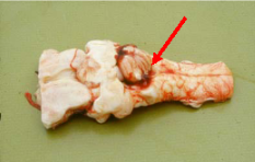
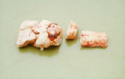
Materials
Equipment
Obex section(s) stained with haematoxylin eosin (H&E).
Obex section(s) labelled immunohistochemically for prion protein (PrP) [for an example of an immunohistochemical protocol, see the document: ‘Fixation, tissue processing, histology and immunohistochemistry procedures for diagnosis of animal TSE (BSE, scrapie, atypical scrapie, CWD)’.
Relevant control material, prepared at the same time as the diagnostic sample(s).
Light microscope with low, medium, and high-power objectives.
Method
Check that the identification on each labelled slide corresponds with that recorded on the accompanying paperwork.
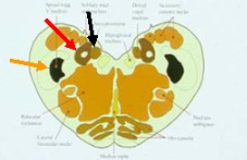
Examine each section (and the relevant controls) methodically using standard histopathological practices. The major target sites at the level of the obex include the solitary tract nucleus, the dorsal nucleus of the vagus nerve, the spinal tract nucleus of the trigeminal nerve and the olivary nuclei.
The following guidelines will aid interpretation:
Diagnostic outcomes
1. Positive (histopathology):
The presence of characteristic vacuolation (spongiform change) affecting the neuropil with an anatomically precise and usually bilaterally symmetrical distribution. Vacuolation in a single target site is sufficient for histopathological confirmation. Usual target sites in bovine are the solitary tract nucleus and the nucleus of the trigeminal tract, and in ovine the dorsal nucleus of the vagus nerve. In atypical scrapie the obex areas that are usually affected are the nucleus of the spinal tract of the trigeminal nerve and the inferior cerebellar peduncle. The area that is consistently affected is the cerebellum and it is for this reason that cerebellum is sampled.
A positive bovine case should usually show more than three neuropil vacuoles per target site. (Wells et al., 1989). It is not possible to be this precise for ovine suspects due to the occurrence of incidental neuronal vacuolation in some sheep (Zlotnik & Rennie, 1957).
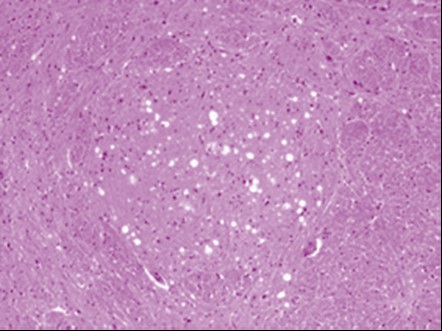
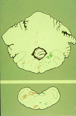
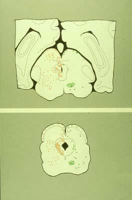
Figure 4: Brainstem sites at which neuropil vacuolation (red dots) and neuronal vacuolation (green dots) can be seen in classical BSE. Generally, these lesions are bilaterally symmetrical. They are represented unilaterally in these diagrams for clarity.
2. Positive (immunohistochemistry) 1, 2
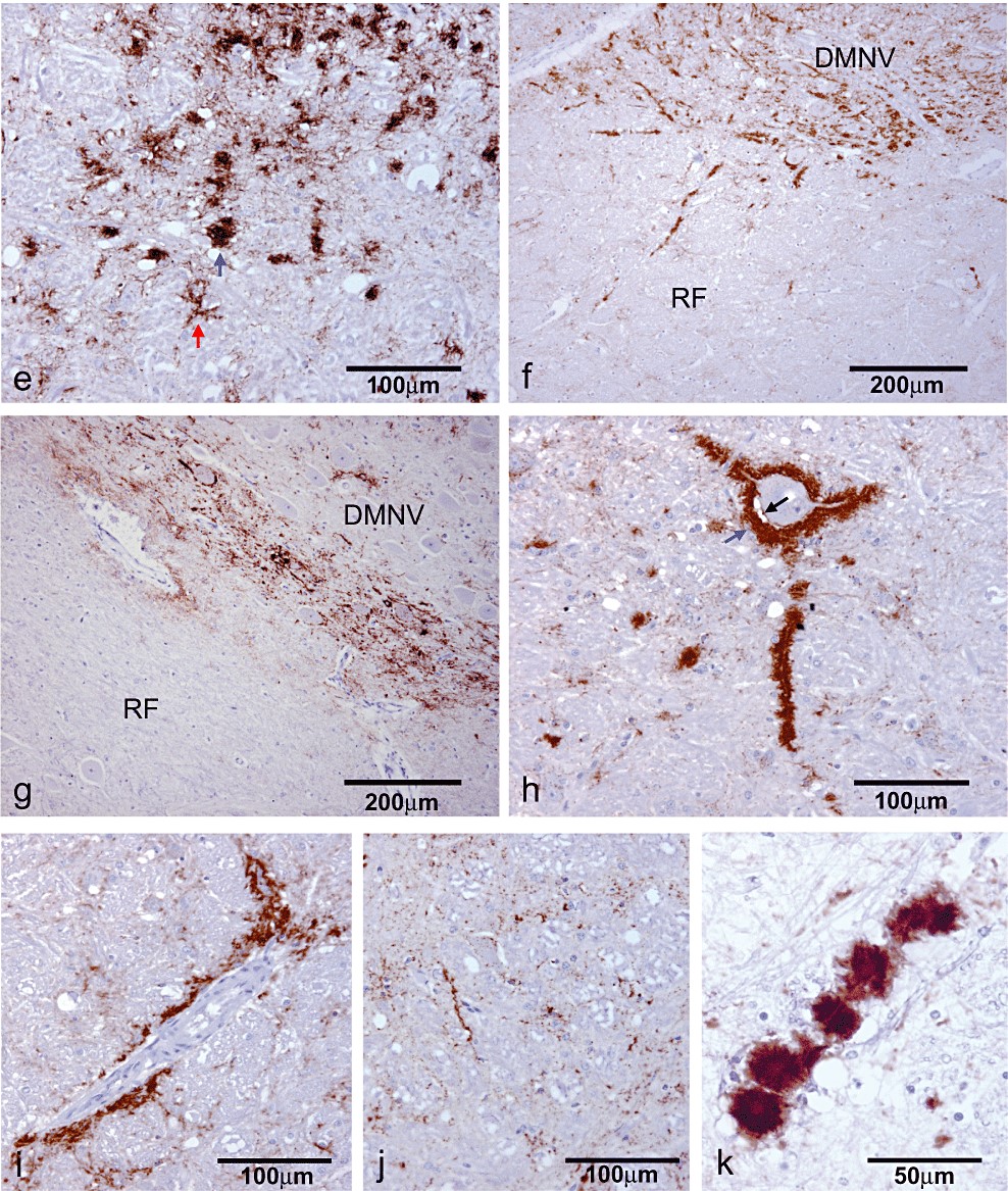
PrPSc immunolabelling is usually found co-localised with disease-specific vacuolation and may also be observed in the absence of spongiform changes.
Non-specific immunolabelling can occur, and it is important to have an awareness of the kinds of immunolabelling which your technique in your laboratory may produce in known negative control animals, so that disease-specific immunolabelling can be identified and correctly interpreted. Examples of the kinds of non-specific labelling which might be encountered can be found in Simmons et al 3.
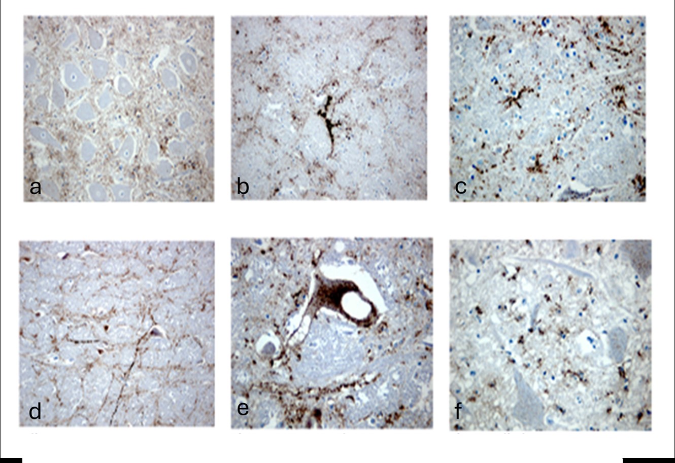
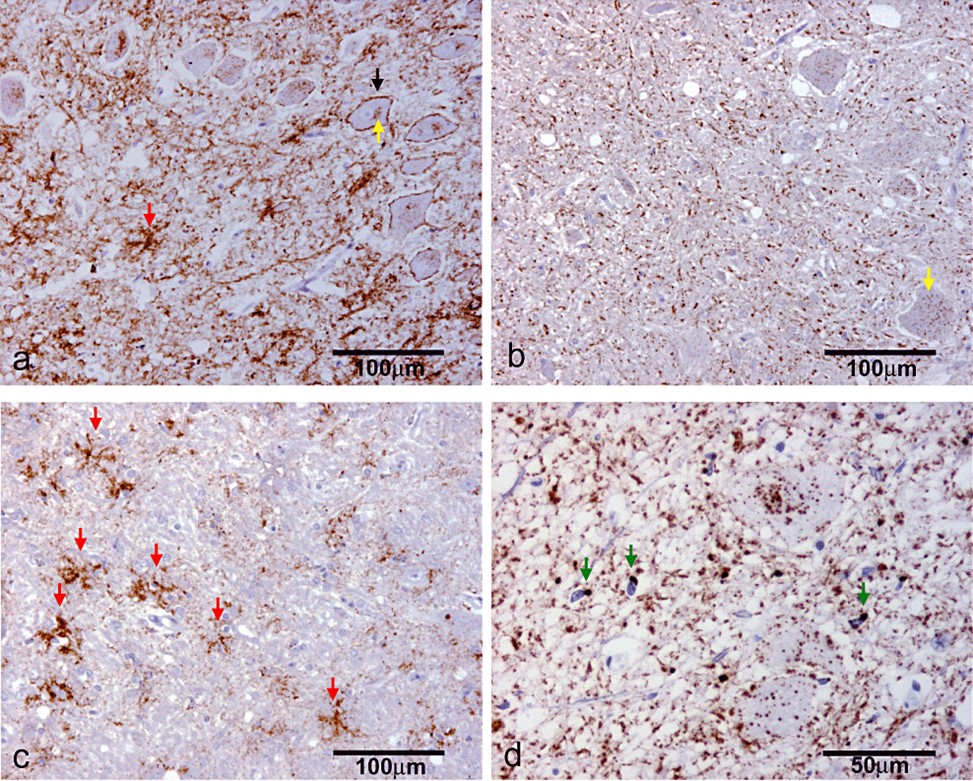
Specific immunolabelling can be found in many different forms, depending on factors such as host species, genotype, and TSE strain.
Some examples are shown in Figures 6 and 7, to illustrate this diversity.
3. Inconclusive (histopathology)
If the target areas are present but contain insufficient neuropil vacuolation to allow unequivocal confirmation by above criteria.
4. Inconclusive (immunohistochemistry)
If the target areas are present, and there is equivocal immunolabelling confined to the TSE neuropil target areas.
5. Unsuitable (histopathology)
If the submitted sample is inadequate, for example due to either poor (or no) representation of target sites, or there is confounding autolysis or other artefactual changes.
This classification may also be used in cases where there are lesions indicative of an alternative neuropathological diagnosis (eg inflammation) which prevent assessment of TSE target sites for the presence or absence of specific spongiform change.
6. Unsuitable (immunohistochemistry)
If there is an absence of appropriate target sites, or the run encountered technical failures eg failure of the positive run control.
7. Negative (histopathology)
A case can be classified as negative if there is an absence of characteristic neuropil vacuolation in sections where the target areas can be confidently identified and assessed.
This classification can also be used in cases presenting with lesions indicative of an alternative neuropathological diagnosis provided adequate assessment of target areas is possible.
8. Negative (immunohistochemistry)
If the target areas are present, and there is an absence of immunolabelling in them. An appropriate positive control must also be present for that technical run.
Atypical TSE
More recently, atypical forms of TSE in both cattle and sheep have been reported. There have also been concerns regarding the possibility of BSE arising in the small ruminant population. As mentioned in the introduction, formal classification of positive isolates is currently performed using discriminatory Western blotting techniques, but there are some variations in the pathology and immunopathology of such cases which should indicate or reinforce the classification of these variants.
The basic principles of confirmation remain the same, but it may be necessary to look at additional areas, particularly the cerebellum in sheep, in order to conclusively confirm or refute a suspicion of atypical scrapie in sheep.
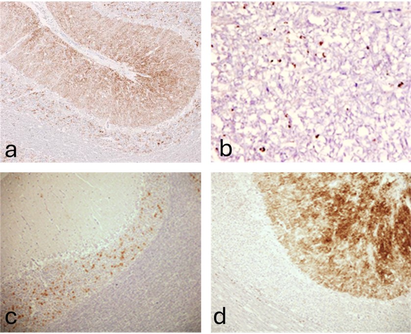
Atypical scrapie in sheep 4 presents with minimal vacuolation or PrP accumulation in the brainstem. However, abundant PrP labelling is generally present throughout the cerebellum, with or without accompanying vacuolation.
Fine particulate forms of PrP deposition are most prominent, with some aggregates particularly in the rostral midbrain and thalamic regions. No intraneuronal intraglial or glial forms are detectable. Vacuolation is most prominent in the basal ganglia and cerebral cortex.
In some, but not all, cases distinctive globular labelling is present in the white matter.
Atypical (H-BSE and L-BSE) in cattle 5 is classified on the basis of the Western blot characteristics of the isolates (see the 2-blot protocol for PrPres typing in BSE from cattle, mentioned earlier in this document).
Confirmation that these cases are positive is straightforward, since both PrPSc accumulation and vacuolation occur in the obex.
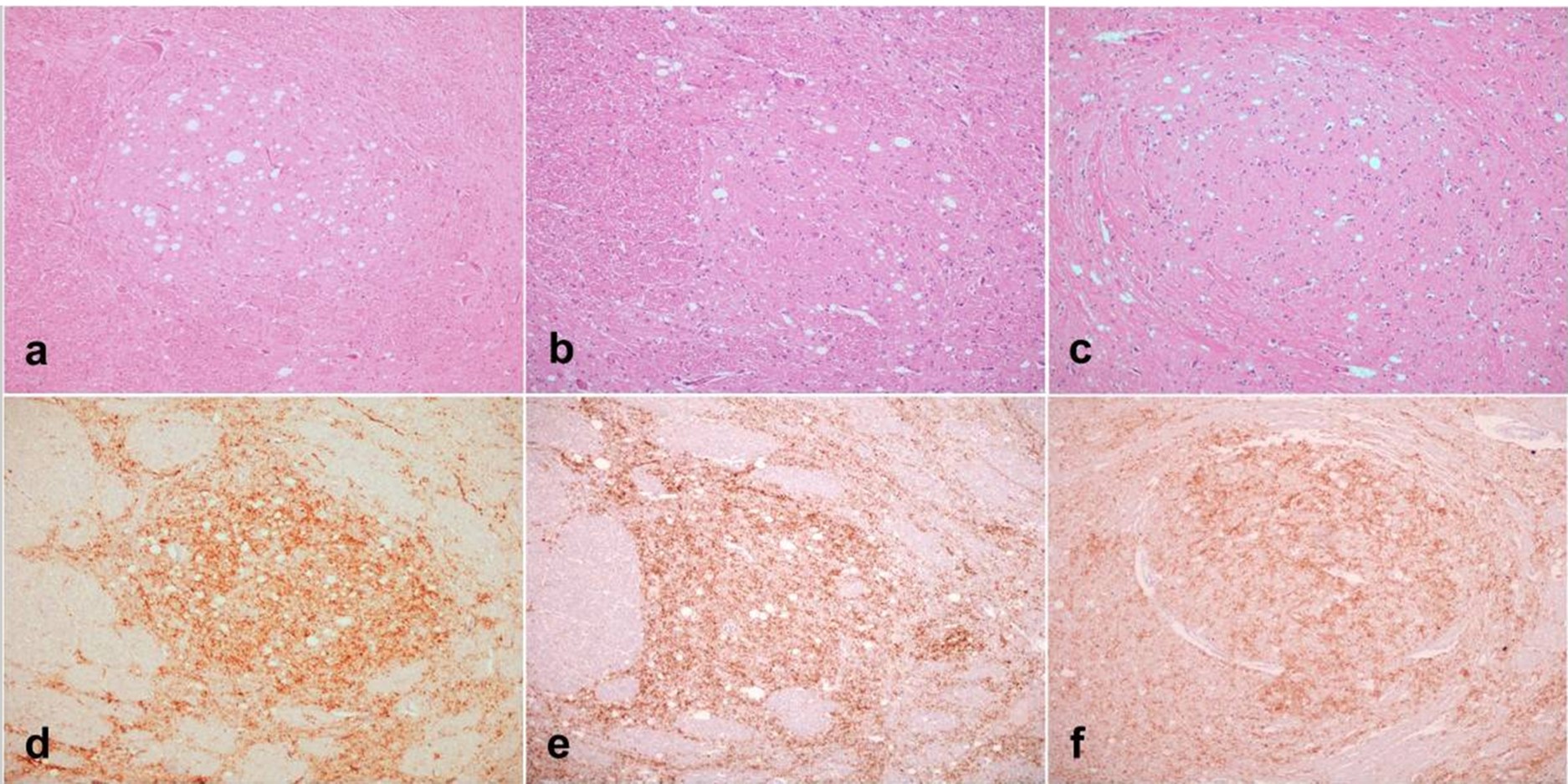
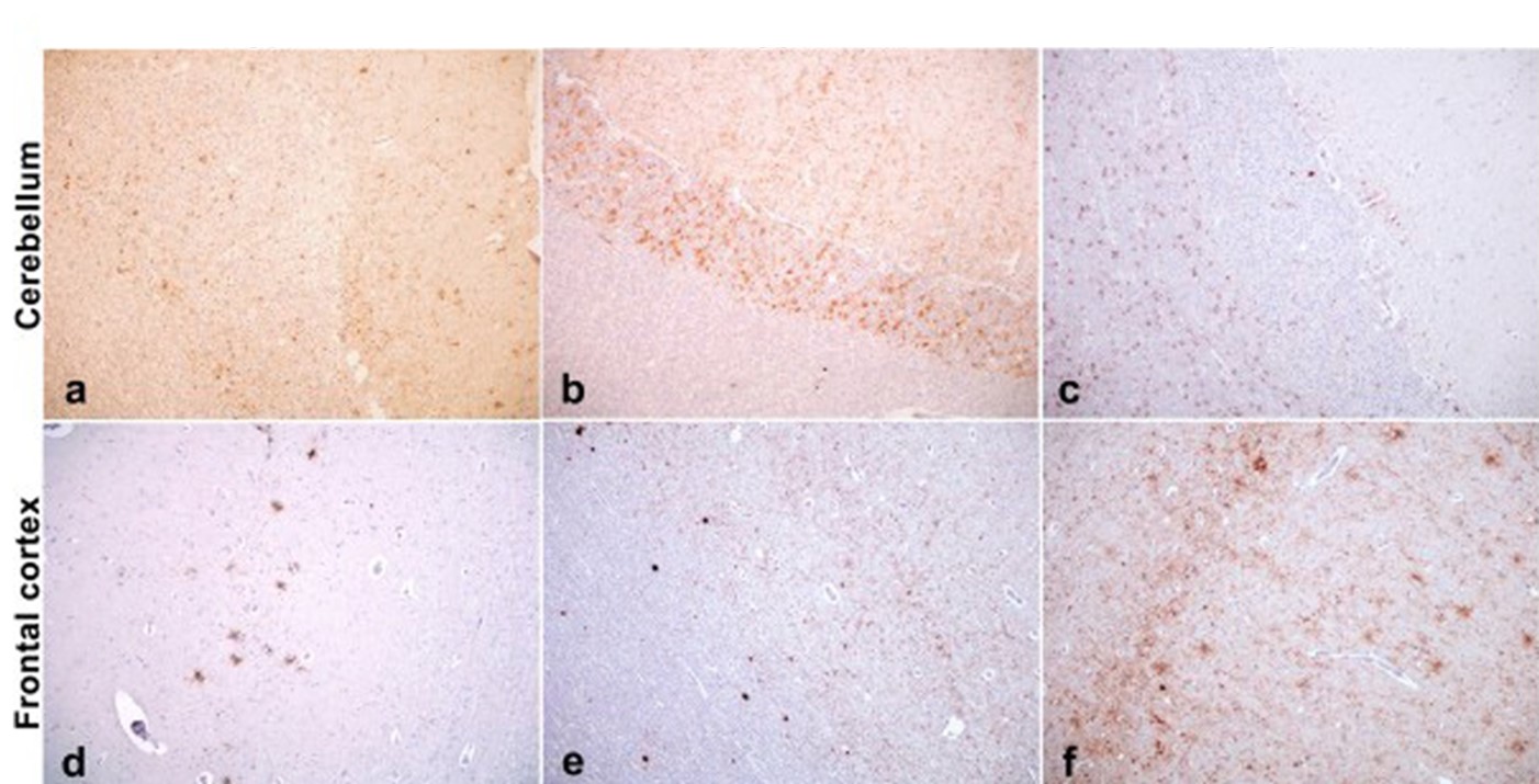
However, the differences between atypical and classical BSE (C-BSE) are subtle in this region, so wherever possible other brain areas (at least the cerebellum7, Figure 10) should also be collected to provide more robust evidence in support of the formal WB classification.
1 Casalone, C., Caramelli, M., Crescio, MI., Spencer, YI and Simmons, MM (2006) BSE immunohistochemical pattern: a comparison between English and Italian cases. Acta Neuropathologica (Berl). 2006 May; 111(5):444-9.2
2 Simmons MM, Spiropoulos J, Webb P, Spencer YI, Czub S, Mueller R, Davis A, Arnold ME, Marsh S, SAC Hawkins, Cooper JA, Konold T, Wells GAH. (2011) Experimental classical bovine spongiform encephalopathy: definition and progression of neural PrP immunolabeling in relation to diagnosis and disease controls. Vet Pathol 48: 948-963
3 Simmons MM, Spiropoulos J, Webb P, Spencer YI, Czub S, Mueller R, Davis A, Arnold ME, Marsh S, SAC Hawkins, Cooper JA, Konold T, Wells GAH. (2011) Experimental classical bovine spongiform encephalopathy: definition and progression of neural PrP immunolabeling in relation to diagnosis and disease controls. Vet Pathol 48: 948-963
4 Moore SJ, Simmons MM, Chaplin MJ and Spiropoulos J (2008) Neuroanatomical distribution of abnormal prion protein in naturally occurring atypical scrapie cases in Great Britain. Acta Neuropathologica 116, 547-59
5 Konold T, Bone GE, Clifford D, Chaplin MJ, Cawthraw S, Stack MJ, Simmons MM (2012) Experimental H-type and L-type bovine spongiform encephalopathy in cattle: observation of two clinical syndromes and diagnostic challenges. BMC Vet Res 8:22