
Clinical signs of transmissible spongiform encephalopathies (TSEs) in sheep
Introduction
The historical protocol to distinguish different strains of scrapie uses mice, which are inoculated with brain tissue from sheep affected by transmissible spongiform encephalopathies (TSEs)11. More recently, strains have been characterised by the regional distribution of disease-associated prion protein (PrPd) in the brain using immunohistochemistry and by the characteristic protein pattern of tissue samples using Western immunoblotting methods27. This has led to a new classification system of scrapie, which distinguishes classical scrapie as it has been known for centuries from atypical scrapie: lesions of the latter are found in older animals with a genotype associated with a lower risk of classical scrapie20. In addition, sheep can be infected experimentally with the agent associated with bovine spongiform encephalopathy (BSE), the characteristics of which can be distinguished from scrapie by immunohistochemical and Western immunoblot examination of the brain. No natural BSE infection in sheep has yet been reported74. Although there may be overlap in the clinical picture of these TSEs in sheep, each type is described separately in the text below.
Classical scrapie
Case characteristics
Most clinically affected classical scrapie cases are between two and five years of age but clinical disease may occur in younger and older animals. There is a report of a case diagnosed as early as 7 months of age65 although it is not known whether this sheep was clinically affected, since pre-clinical infection is often detected in field outbreaks where the affected flock is further investigated following culling10. Experimental infection of sheep has resulted in the onset of clinical signs from 7 months post inoculation21.
The susceptibility or resistance to scrapie is determined by the prion protein (PrP) gene and appears to be independent of sex and breed. Animals with the highest susceptibility to classical scrapie have a genotype that encodes QQ (glutamine) at codon 171 (VV136RR154QQ171 or AA136RR154QQ171 in breeds that usually do not carry the VRQ allele), whilst animals with the PrP genotype AA136RR154RR171 are genetically most resistant to classical scrapie38.
Three codons of the PrP gene are of main importance for scrapie susceptibility: 136 (A = alanine or V = valine), 154 (A = arginine or H = histidine) and 171 (Q = glutamine or R = arginine).
Affected flocks usually experience only small numbers of clinical cases in any year, sometimes only single cases but this is obviously dependent on the genotype distribution within the flock. More explosive outbreaks have been reported, but are relatively rare. Most cases have an insidious onset and progress slowly over several weeks. In some cases, clinical signs may be observed for up to 7 months. Early clinical signs may not be noticed, especially in flocks that are not closely monitored. It is unclear if acute or peracute scrapie cases exist that do not display premonitory signs of illness. In one survey, 16% of confirmed scrapie cases were found dead without exhibiting signs before death17. Scrapie cases without apparent signs of scrapie but widespread vacuolation in the brain have also been diagnosed in abattoir surveys.
Clinical signs
Early clinical signs can be fairly unspecific and are thus most likely to be observed by someone familiar with the animal. Farmers with experience of the disease may suspect an early case of scrapie in a flock in which the disease is endemic before neurological signs are evident. These early clinical signs may include weight loss and subtle behavioural changes.
Changes in mental status, behaviour and activity
Changes in mental status, behaviour and activity are often the earliest clinical signs to be noticed. Animals may separate themselves from the rest of the flock and may become more flighty when approached and thus more difficult to catch. Affected sheep may also appear hyper-alert, may become more excitable and some may display exaggerated responses to external stimuli, such as sudden movements or noises, e.g. created by a passing tractor or a hand clap. When left undisturbed, scrapie cases may appear somnolent or dull with a vacant gaze and lowered head. This may become more obvious and permanent in advanced scrapie cases but may only be episodic in the early stage and thus only detectable following prolonged observations.
Social interaction seems to be reduced in scrapie-affected sheep32 and cases tend to spend more time lying down than healthy sheep: they are usually more inactive during these periods. Teeth grinding is frequent in scrapie cases and occurs either spontaneously during periods of inactivity, whilst being handled, or in combination with lip licking.
Video 1
This 2-year-old Swaledale ewe (ARQ/VRQ) is licking its lips excessively, which is sometimes accompanied by teeth grinding.
Appetite is usually not affected until the very late stage of the disease although it has been reported that scrapie-affected sheep spend less time feeding on hay. In addition, mastication and bolus regurgitation may be reduced in sheep with scrapie2;32. Abnormal drinking patterns have also been reported in the early stages of scrapie. Sheep with scrapie may drink more frequently but take only small amounts each time56.
Frequent urination (pollakiuria) of small amounts of urine may be observed in some scrapie cases, which has been attributed to hyperactive function of the detrusor urinae muscle and variation in the urethral tone12.
Pruritic activities
Grooming behaviour seems to be increased in most sheep with scrapie. Scrapie cases spend more time rubbing on objects (e.g. fences), scratching, licking and self-nibbling. In the later stages, rubbing may be accompanied by lip licking and smacking, which gives the sheep an expression of satisfaction. These pruritic activities are however often only displayed when the animal is undisturbed or accustomed to the presence of an observer.
Video 2
This 4-year-old Swaledale ewe (VRQ/VRQ) displays pruritic behaviour, such as scratching her belly with her hind leg and movements of lip and tongue when she rubs her back briefly on the hay rack.
Although pruritus is a classical feature of scrapie, some cases may not display this sign, a feature which led to the classification of an 'ataxia type of scrapie'54 or a 'paralytic form of scrapie'39.
Scratch test
This test is used to assess the sheep’s response to scratching its back. The scratch test sites are displayed in figure 1. Test sites should be scratched repeatedly (at least 3 times) and with a pause between each stimulus.
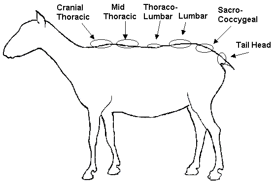
A repeatable, stereotypical response is a positive scratch test response. The test requires a fairly quiet and immobile animal since a response may not be detected in excited animals. Ideally, handlers should stand next to the sheep and use their legs to ‘press’ the animal against a wall (see figure 2).
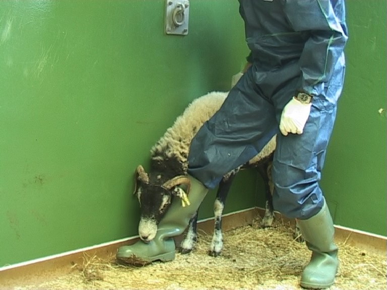
Most scrapie cases respond to scratching of the back with lip licking and nibbling (similar to the self-induced response; frequently but not always also with rapid extrusions of the tongue), which has been called the nibble reflex).
Video 3
This 5-year-old Texel ewe (ARH/ARQ) licks its lips when the sacral area of the back is scratched.
In advanced cases of scrapie, lip smacking may be elicited by simply touching the animal’s back. Some cases, however, respond to the scratch test with only rhythmical head or body movements (curling, flexion or extension of the head and neck, or body swings) without clear nibbling lip movements although this response is still considered a positive scratch test response.
Video 4
This 5-year-old Swaledale ewe (ARQ/VRQ) responds to scratching of the midthoracic area of the back with head movements. This is accompanied by lip movements when the back is scratched more caudally.
Most pruritic cases will be detected if animals are observed for signs of excessive grooming as some animals may not respond to the scratch test. In a study of 162 sheep with scrapie, 32 (20%) did not respond to the scratch test, even though they were pruritic when observed for a few hours. Conversely, 18 sheep (11%) had a positive scratch test without displaying any other activity indicating pruritus; only 6 sheep (4%) did not display either sign 40.
A positive scratch response was reportedly more frequent in Irish sheep of the genotypes ARH/ARQ or ARQ/ARQ than in sheep of the genotype ARQ/VRQ 33.
The scratch test is not specific for scrapie since pruritic skin conditions, e.g. mange, can cause the same response.
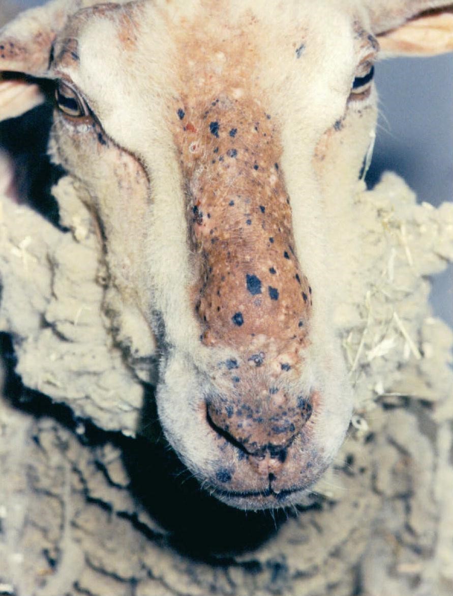
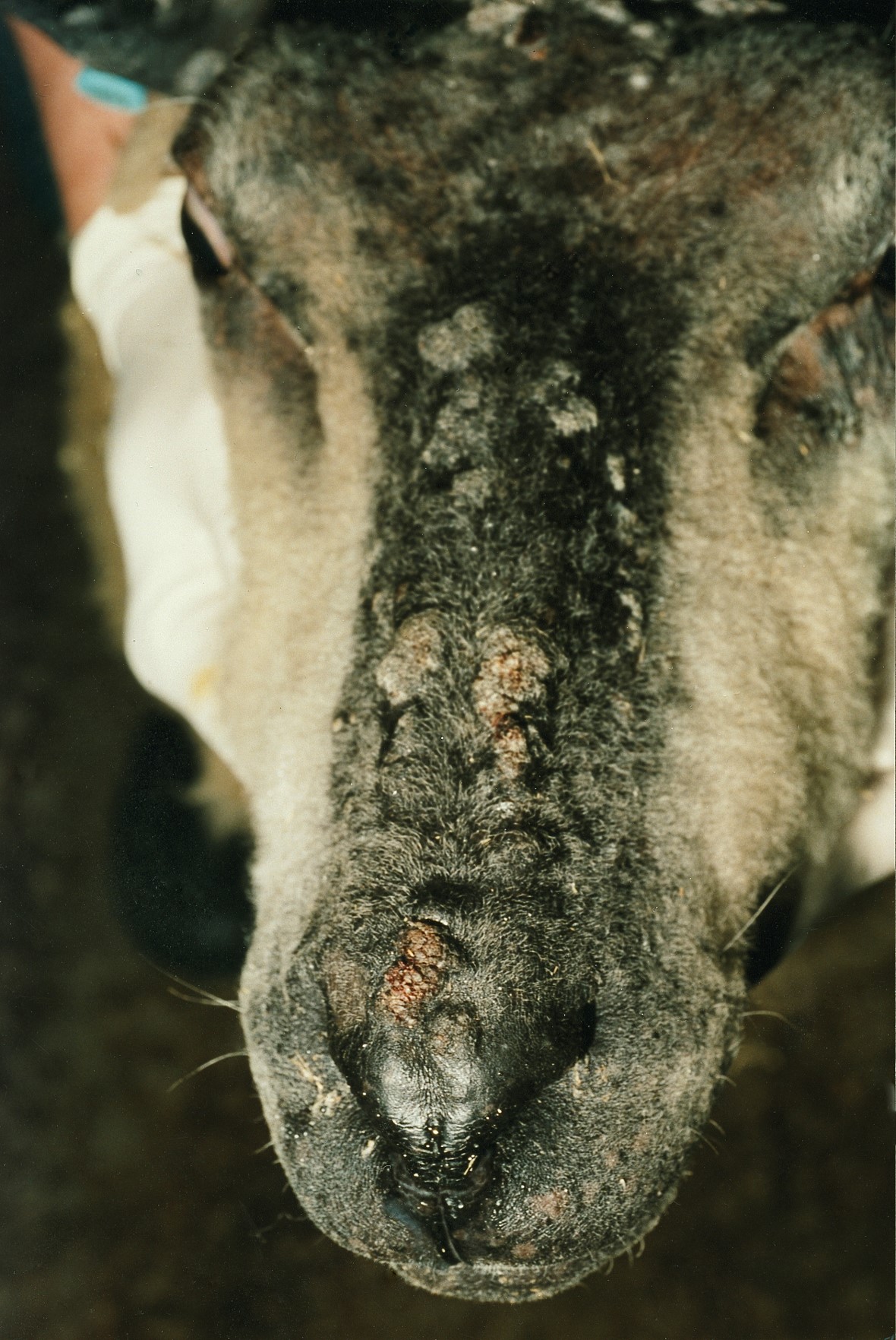
Intense nibbling, rubbing and scratching will eventually lead to discolouration and loss of wool as well as hyperpigmentation and lesions of the skin. Skin lesions initially present as superficial abrasions, excoriation with crusts if allowed to heal and lichenification. Such lesions may become secondarily infected by skin pathogens (e.g. Staphylococcus aureus, Dermatophilus congolensis 40), which results in the formation of nodules and pustules. It is also possible to isolate parapox-virus (orf) from these skin lesions. Figs. 3-4 show skin alterations observed in scrapie cases.
Postural changes
Scrapie cases may stand with their hind limbs placed in an abnormal position (e.g. wide-based stance); they may also stand with their hind quarters low and their hind limbs partially flexed in a ‘crouching’ posture 33;39.
Gait changes
Gait abnormalities are usually observed later in the course of the disease in cases that present with the ‘classical’ signs – behavioural changes, pruritus and weight loss. Gait deficits are often enhanced upon exercise, on turns and when going over steps. The gait may initially have a stiff appearance. Ataxia, characterised by a wobbly, uncoordinated gait, is usually more frequent in the hind limbs. Bunny hopping (hopping simultaneously with both hind limbs) may also be observed when sheep are made to run. A high stepping, dressage-type gait (hypermetria) may be present, which led to its German name ‘Traberkrankheit’ (trotting disease). Severe gait deficits ultimately result in difficulty getting up and eventual recumbency.
Video 5
These three 2-year-old crossbred ewes show various degrees of ataxia and limb weakness: as they walk off the straw-covered floor gait abnormalities are mildest in the first ewe (marked wool loss, ARQ/ARQ), more severe in the second ewe (ARH/ARQ) and worst in the third ewe (ARQ/VRQ), in which the fore limbs are also affected.
Involuntary movements
Scrapie cases frequently exhibit a tremor of the head, which may be more evident when sheep are stressed, e.g. during or after handling. This may later progress to generalised (whole body) tremors. Some cases may display myoclonic muscular contractions 26;51. Electromyographic studies have shown that tremors consist of two components that occur asynchronously: tetanic contractions of skeletal muscles and trembling of skin muscles 39.
Video 6
This 7-year-old Charollais crossbred ewe (AHQ/ARQ) exhibits a fine head tremor at rest.
Seizures may be seen in some cases of scrapie 48 and may be precipitated by handling and excitement 57.
Collapsing episodes, which may be a form of cataplexy-narcolepsy, have been occasionally observed. Sheep may suddenly collapse for up to several minutes when stressed (e.g. during handling or when chased by a dog) and then regain their feet unaided 63;73.
Video 7
This 3-year-old Welsh Mountain ewe (VRQ/VRQ) presents with collapsing episodes suggestive of catalepsy: She displays a flaccid paralysis without loss of consciousness since she still (over-)reacts to testing the menace response.
Weight loss and loss of body condition
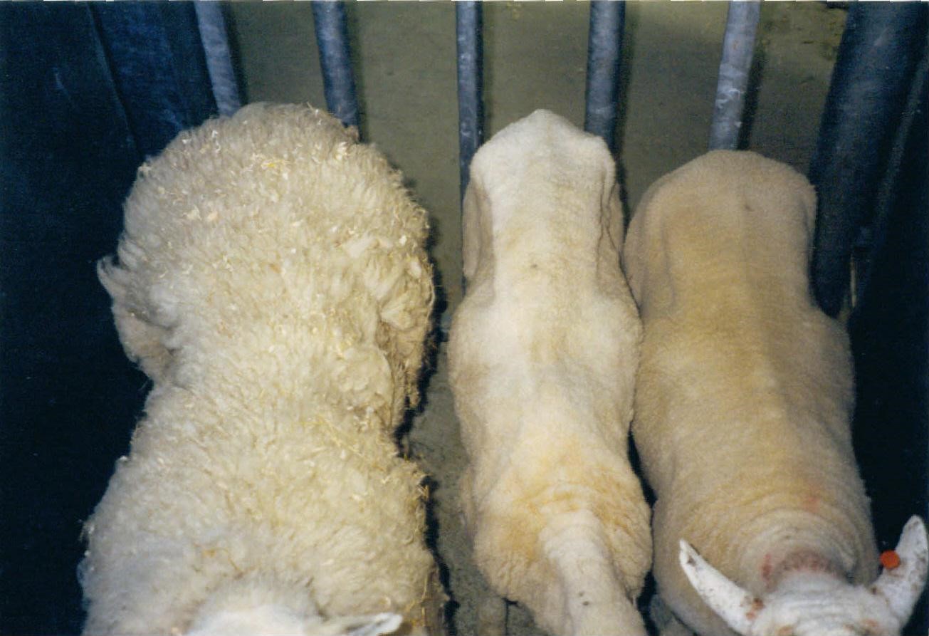
Loss of weight and body condition (see Fig. 5) despite an apparently maintained appetite is present in the majority of scrapie cases and can be a very early but unspecific clinical sign 82. Weight loss may progress slowly to emaciation in advanced scrapie cases 33.
Other miscellaneous clinical signs
Dribbling of ruminal fluid and salivation
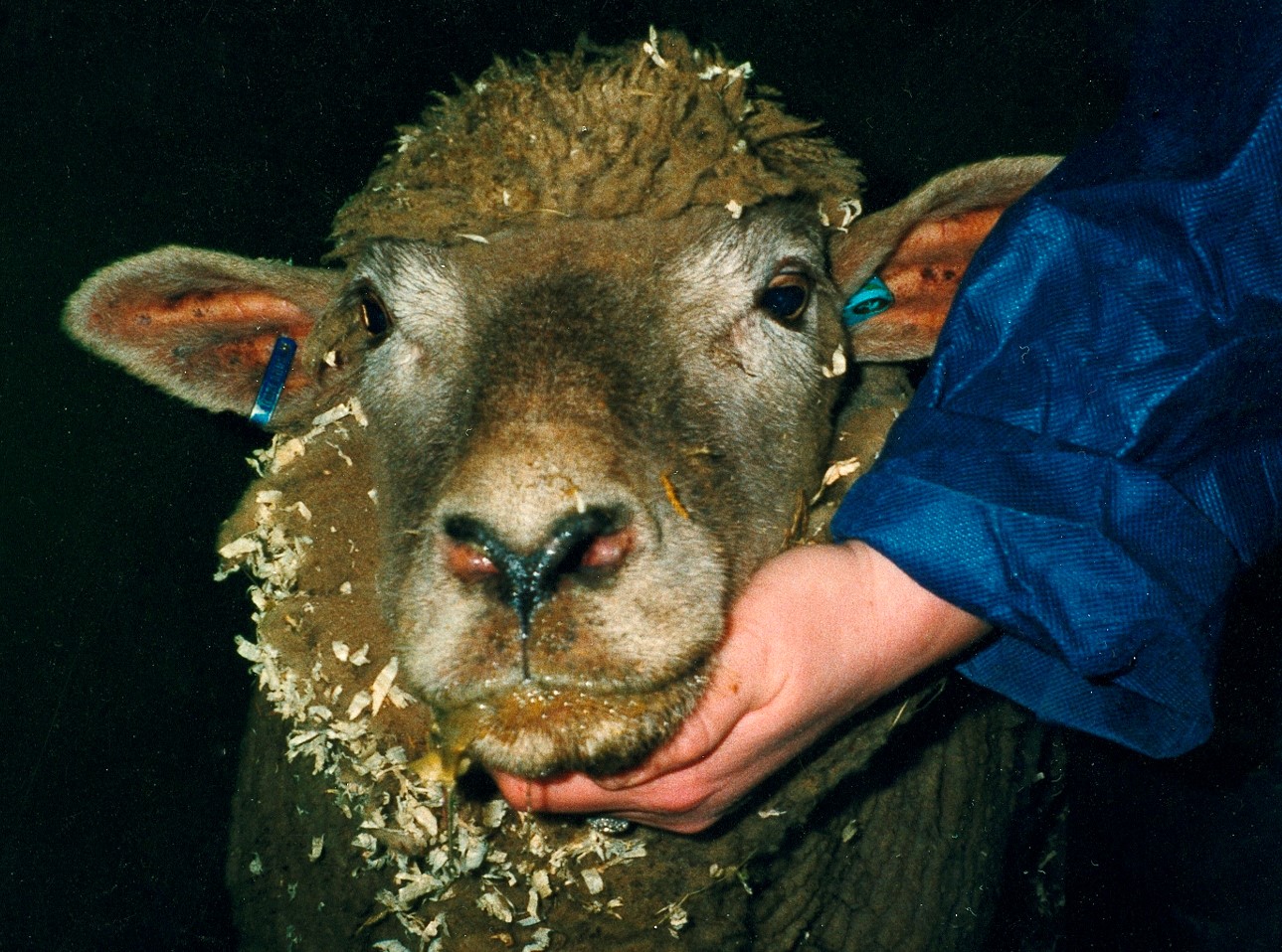
Drooling saliva may be present in some cases 48. Dribbling of ruminal fluid with consequent staining of the wool around the nostrils, mouth and chin has been observed occasionally in scrapie cases 33 (see Fig. 6): this may be the result of swallowing deficits 55.
Ocular abnormalities
There are occasional reports of scrapie cases running into objects. Although this may be attributable to panic reactions in excitable animals, visual impairment has nevertheless been reported infrequently 30;79;81. Other rare abnormalities include nystagmus 55;57, an absent menace response 4;33;75;82 or a reduced pupillary light reflex 39;82.
Video 8
This 3-year-old Suffolk ewe (ARQ/ARQ) does not display a menace response. This may be caused by dysfunction of cerebellar pathways, because the animal had a normal blink reflex and was able to leave and re-enter her pen and walk along the corridor without any evidence of visual impairment.
Hyper-reflective oval, blister-like areas throughout the tapetum lucidum were observed in 2 sheep with signs of visual impairment 4 but no appreciable fundoscopic changes were observed in 17 scrapie-affected sheep despite evidence of functional and morphological changes in the retina 60.
Hypoaesthesia
Loss of sensation (hypoaesthesia or even anaesthesia) in the distal parts of the limbs has been observed in very early cases of scrapie whilst loss of facial sensitivity may be observed in the late stages of the disease 82.
Cardiac and respiratory abnormalities
Autonomic disturbances (cardiac arrhythmia, accompanied by tachycardia, bradycardia and intolerance to exercise) have been reported 7;42;82 although abnormalities in heart rate and rhythm were not observed in a separate study 33. Heart rate variability has been studied as an aid to the diagnosis of scrapie but its usefulness is controversial 28;42.
Scrapie cases may display an increased respiratory rate characterised by panting when stressed, e.g. after handling, or may show increased respiratory sounds. Scrapie has been diagnosed in a ewe that only displayed signs of respiratory disease 85. It is important to note that sheep affected with scrapie may suffer from secondary infections, e.g. clostridial infections, pneumonia 9;35.
Ruminal impaction
Gastrointestinal stasis has been reported in some scrapie cases 63;64.
Milk production
Hypogalactia and difficulty in milking is frequent in sheep that are used for milk production 14.
Biochemical markers in body fluids
Some changes in blood neurotransmitters, hormones, metabolites, metals or immunoglobulins have been found in scrapie cases, but it has so far not resulted in a test that would allow scrapie diagnosis in live animals. These changes included decreased blood serotonin 16, increased plasma 20beta-dihydrocortisol and urinary creatinine 58, increased concentrations of serum IgG 18, plasma growth hormone 83, blood copper and blood manganese 34. Alterations in energy metabolism as determined by changes in the plasma concentrations of citrate, lactate and 3-hydroxybutyrate have also been reported 15.
Cerebrospinal fluid analysis was unable to distinguish scrapie cases from healthy sheep 63. Other markers associated with neuronal damage in the brain, such as 14-3-3 protein detection in cerebrospinal fluid, have not been found useful for scrapie diagnosis 80. An increase in acute phase proteins (ceruloplasmin, haptoglobin and serum amyloid A) in serum of clinically affected sheep with natural and experimental scrapie has been reported 52 but such increases are likely to be too nonspecific for a diagnostic test.
Recent research has concentrated on the detection of disease-associated prion protein in blood/ serum 3;61;77;78 or other fluids/ excretions 29;49;76. Although the results are promising, an evaluation of their usefulness for routine diagnosis using a large dataset has not been conducted.
Electrodiagnostics
Abnormal electroencephalograms were obtained from a few sheep with scrapie 39;75. Two confirmed scrapie cases had reduced amplitudes in brainstem auditory evoked potentials and flash evoked visual potentials 75; in a larger study, 23% of 182 clinically affected sheep with TSEs presented with abnormal auditory evoked potentials 47. Alterations in the electroretinogram of scrapie-affected sheep, characterised by reduced wave amplitudes, have also been described 60;72. Electrodiagnostic tests are expensive, time-consuming and thus may not be of practical value, but they may be useful to demonstrate lesions in certain neuroanatomical structures. However, they have been applied to only a very limited number of animals, and it is not known if these tests detect scrapie in sheep of various breeds and genotypes infected with different strains.
Fluorescence spectroscopy of the retina was proposed as another eye-based test for the diagnosis of scrapie although it has not been applied to live animals 1.
Clinical diagnostic criteria
Based on the observation that scrapie cases display pruritus and ataxia, the absence of a nibble reflex alone or in combination with absent proprioceptive deficits was judged to be highly indicative of a scrapie-negative sheep on farms with reported cases of scrapie 19. These criteria were proposed as an aid for scrapie surveillance in Italy.
A scoring system has been proposed to assess the clinical progression in classical scrapie based on the presence of eleven specific clinical signs: hypoaesthesia in the limbs, body condition score of less than 3 (out of a maximum of 5 for an obese sheep) and alteration of mental status were given a value of 1, hyporeflexia in limbs, cardiac arrhythmia, pruritus/ wool loss and nibble reflex had a value of 2 and head tremor, hyperexcitability, ataxia/ gait abnormalities and teeth grinding had a value of 3 82. A different value was applied to different clinical signs to take into account the fact that certain clinical signs like teeth grinding, ataxia and tremor occur later in the disease. By adding the values of each sign if present the authors determined that a sum of 1-4 represented an early stage, a sum of 5-13 an intermediate stage and a sum of 14-23 a late stage of the disease. This scoring system has only been evaluated in Rasa aragonesa sheep with an ARQ/ARQ genotype.
Both clinical protocols require a close examination of sheep and familiarisation with neurological assessments of animals, which cannot always be expected from the keeper of sheep. It is important that farmers are able to recognise signs of disease, which can be variable as mentioned above, in order to report the disease to the local veterinary authority. The proposed clinical protocols may be useful to confirm the diagnosis of a suspect scrapie case reported by the farmer.
A short clinical examination protocol (see article and video) has been proposed to check for specific clinical signs associated with pruritic and non-pruritic forms of TSEs in sheep, which includes assessment of behaviour, vision (by testing of the menace response), pruritus (by testing the response to scratching) and movement (with and without blindfolding). The combined occurrence of two or more signs such as a repeatable response to scratching, tremor, abnormal behaviour, circling, collapsing episodes, ataxia or an absent menace response were considered highly suggestive of scrapie 45. When this examination protocol was applied to over 1,000 sheep, including sheep naturally or experimentally exposed to a classical, atypical scrapie or bovine spongiform encephalopathy agent, the main clinical markers to predict TSEs were incoordination and a positive scratch test 46.
CH1641 strain
The CH1641 scrapie strain was originally derived from a Cheviot sheep with naturally occurring scrapie, which has been subpassaged in sheep for further characterisation 23. The molecular phenotype has similarities to classical BSE in sheep 36, which makes it diagnostically more challenging. Similar 'CH1641-like' scrapie cases have been described in other natural cases of scrapie in France 5. Sheep experimentally infected with the CH1641 isolate presented with ataxia and infrequently loss of the menace response but were not pruritic and similar clinical signs were observed in a sheep experimentally infected with an atypical scrapie isolate that produced CH1641-like characteristics on pathology, biochemistry and murine bioassay 71. A video showing the clinical signs in this sheep is available within the published manuscript in PLoS One 71.
Atypical scrapie
A new strain of scrapie that caused mainly ataxia in the absence of pruritus has first been described in Norway and was called Nor98. Disease-specific prion protein was found predominantly in cerebellar and cerebral cortices, and the molecular profile by Western immunoblot differed from that usually seen in scrapie (protein band of lower molecular mass than in previous scrapie cases) 8. More of these cases have since been detected in other countries, and the disease was named 'atypical scrapie' to distinguish it from the previously known scrapie disease, now called 'classical scrapie' 20. Atypical scrapie has been confirmed retrospectively in a clinical suspect sheep culled in 1987 84, which suggests that this condition is not new.
Although there is an overlap in the range of PrP genotypes of sheep affected by atypical and classical scrapie, atypical scrapie cases often have a genotype different to classical scrapie cases: such cases are rarely VRQ homo- or heterozygous but atypical scrapie has been found in ARR homozygous sheep, which are considered to be highly resistant to classical scrapie 62.
Most cases were detected in fallen stock or abattoir surveys; clinically affected animals have rarely been reported. Affected sheep are usually older, above 3.5 years of age.
Major clinical signs are ataxia and behavioural changes, such as nervousness or anxiety, in addition to loss of weight or condition 8;44;53. More detailed examination may reveal tremor and a deficient menace response 43. Circling and visual impairment have also been observed 66.
Video 9
This 3-year-old crossbred ewe (AHQ/AHQ) was reported as suspect following ante mortem inspection in an abattoir. It is circling clockwise and displays a high-stepping (hypermetric) hind limb gait. This sheep also had an absent menace response. Note the wool loss on the back but it did not respond to the scratch test and was not observed to be pruritic (the origin of the wool loss was not further investigated).
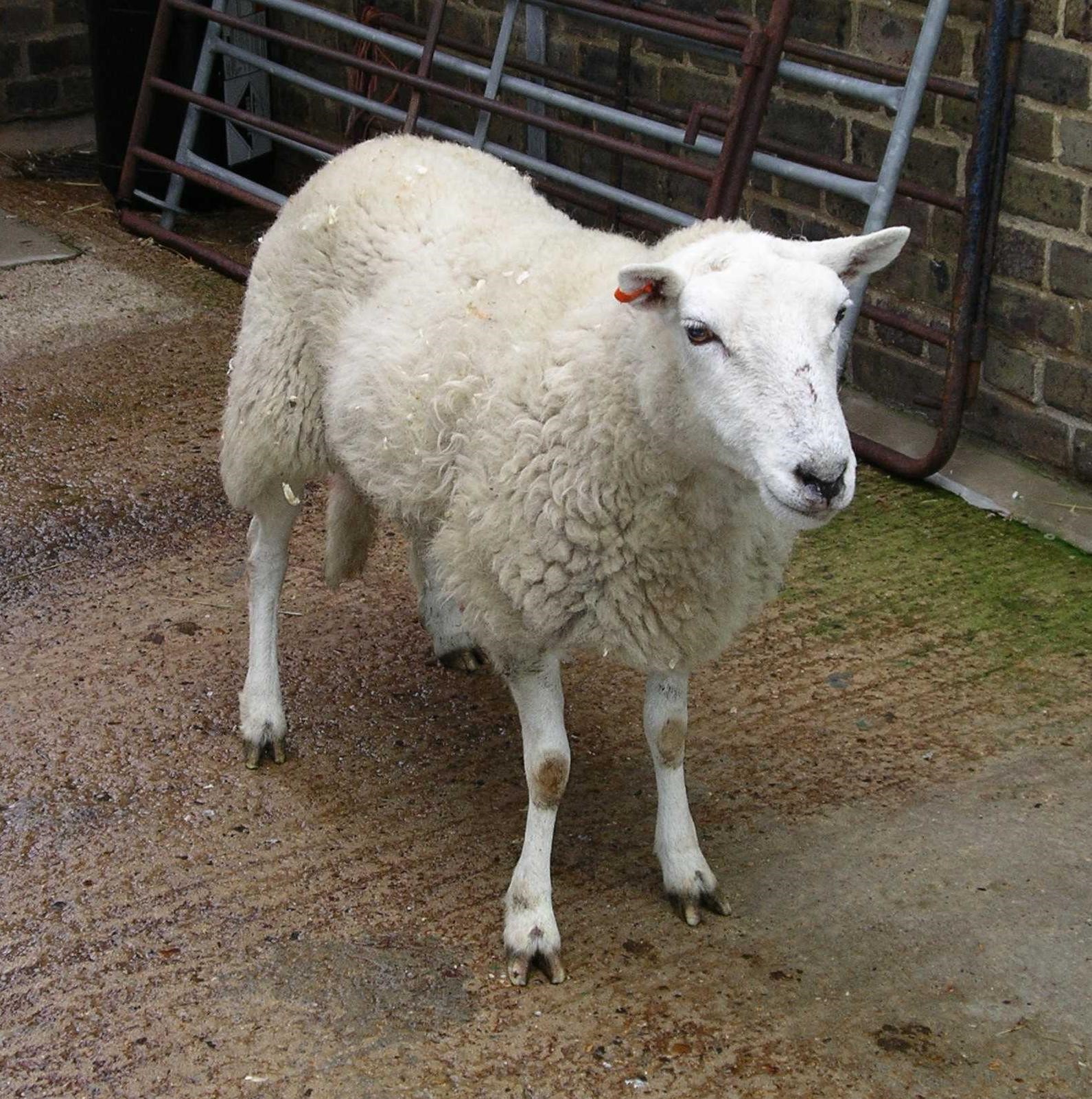
Pruritus or physical changes suggestive of pruritus, such as wool loss, are usually absent 22 (see Fig. 7) although a response (rhythmical head and body movements) to scratching of the back has been reported in one case 43.
Overall, there seem to be similarities to the non-pruritic form of classical scrapie, which may occur in genotypes (ARQ/ARQ, ARQ/ARR) that are also associated with atypical scrapie 40. Circling, however, is usually not considered a sign of scrapie and may often lead to the diagnosis of suspect listeriosis or a unilateral space-occupying lesion in the brain.
Experimental oral or intracerebral infection of sheep with atypical scrapie brain homogenate produced signs similar to those reported in naturally affected sheep, such as abnormal behaviour (compulsive circling, confusion), tremor, ataxia or dysmetria, an absent menace response and weight loss but no signs of pruritus 68-70.
More video clips of clinically affected sheep with atypical scrapie can be found in published open-access manuscripts.
Natural disease: BMC Vet Res – article 1 43 and article 2 66.
Experimental disease: BMC Vet Res 68 and Emerg Inf Dis 70.
BSE in sheep
BSE has not yet been identified in naturally infected animals. Clinical signs of sheep with BSE have been described after experimental infection with the BSE agent using various routes of challenge.
Classical BSE
An extreme short clinical course of less than a week was observed in intracerebrally and orally inoculated Cheviot sheep, and the dominant signs were rapidly progressive ataxia and recumbency, whilst evidence of pruritus was observed in only one animal 24.
In a further experiment where Poll Dorset, Cheviot and Suffolk sheep were inoculated intracerebrally or intravenously, the clinical signs were comparable to ‘classical’ scrapie, although pruritus was less frequent and ataxia more frequent when compared to sheep experimentally infected with scrapie. Weight loss was present in only 11% of the animals 37.
The clinical signs after intra-peritoneal inoculation of one sheep were intense pruritus and ataxia that progressed over a period of 3 months 6. Pruritus was indeed a common feature in sheep of different genotypes that were experimentally and naturally (in an experimental flock) infected; the systematic examination and observation of sheep in these studies, which included testing of the scratch test, may have resulted in the detection of more pruritic cases than in previous studies 41.
Video 10
This 26-month-old Poll Dorset wether (ARQ/ARQ), orally inoculated with 0.5 g of ovine BSE brain homogenate from an ARQ/ARQ donor, displays a positive scratch test, characterised by lip licking and upwards movement of the head, which can be elicited by merely light scratching of the back.
A behavioural study of nine ARQ/ARQ sheep intracerebrally infected with the BSE agent confirmed the presence of pruritic activity in all sheep; there was also reduction in feeding activity whilst resting behaviour and the frequency of low head posture increased 13.
Further video clips documenting the clinical signs of BSE in sheep can be found in an open-access article in BMC Vet Res 41.
Overall, the clinical signs in BSE of sheep appeared to be similar to classical scrapie and included behavioural changes (nervousness or dullness, teeth grinding), signs of pruritus (rubbing, scratching, nibbling, fleece changes including alopecia, a positive scratch test), movement disorders (tremor, ataxia) and loss of weight. Pruritus, tested by assessing the sheep’s response to scratching, was one of the earliest signs displayed in affected animals 41.
Thus BSE, if it occurred naturally in sheep, would be indistinguishable from classical scrapie.
Atypical BSE
Intracerebral inoculation of sheep with bovine atypical (L-type) brain homogenate produced clinical disease with two different phenotypes: a pruritic form indistinguishable from classical scrapie or BSE in sheep and a cataplectic form, characterised by sudden collapse with complete loss of muscle tone, often in combination with sleeping episodes (narcolepsy). This cataplectic form was maintained on subpassage in sheep 67. Examples of the pruritic and cataplectic form are presented as video files within the article in Vet Res 67.
Chronic wasting disease in sheep
Intracerebral inoculation of a US isolate of Chronic Wasting Disease from mule deer to sheep produced clinical disease in only one of four sheep, and the clinical signs were scrapie-like with loss of appetite, weight loss, fine-muscle twitching and progressive recumbency 31, whilst isolates from white tail deer did not cause unequivocal neurological signs 25. It is currently not known whether Scandinavian isolates could cause clinical disease in sheep.
Differential diagnosis of scrapie
Classical scrapie may be confused with other neurological diseases or other diseases that cause a progressive loss of condition or pruritus with skin lesions. Classical scrapie usually does not cause neurological signs that are unilateral or focal in distribution. However, the presence of other diseases, e.g. pregnancy toxaemia or various skin diseases, does not rule out the absence of scrapie.
As atypical scrapie cases may present with circling, which is suggestive of an asymmetrical lesion in the brain, diseases causing similar clinical signs should be considered in the differential diagnosis of atypical scrapie, particularly listeriosis and space-occupying lesions affecting one side of the brain (tumour, abscess, parasitic cyst).
Tables 1 to 3 list the main differential diagnoses for scrapie. Diseases that generally occur in young animals (denoted as *) are listed because classical scrapie has been reported in a sheep of less than one year of age. Details on respective diseases can be found in veterinary textbooks 50;59.
Table 1: Weight loss/ cachexia
Aetiology | Name of the disease |
Infectious | Leucosis, paratuberculosis, pseudotuberculosis (internal form), endoparasites |
Nutritional | Protein/ energy malnutrition, cobalt deficiency |
Miscellaneous | Dental disease, abomasal dilation and emptying defect |
Table 2: Skin diseases
Aetiology | Name of the disease |
Infectious | Mange, parasitic dermatosis (e.g. lice infestation), cutaneous myiasis, dermatomycosis, bacterial dermatitis |
Allergic | Allergic dermatitis |
Miscellaneous | Photodermatitis |
Table 3: Neurological diseases
Aetiology | Name of the disease | |
Infectious | Viral encephalomyelitides (Aujeszky’s disease, Borna disease, louping ill, maedi/visna, rabies), bacterial meningoencephalo-myelitides and epidural abscesses (listeriosis) and focal symmetrical encephalomalacia (Clostridium perfringens), fungal meningoencephalo-myelitides (aspergillosis, cryptococcosis – rare), protozoan encephalomyelitis (toxoplasmosis, sarcocystosis) and cysts or parasitic migration (Coenurus cerebralis) | |
Metabolic | Pregnancy toxaemia, hypocalcaemia-hypomagnesaemia-complex, hepatic encephalopathy | |
Congenital/ Anomalies | Ovine congenital progressive muscular dystrophy*, ceroid lipofuscinosis*, cerebellar abiotrophy*, ovine GM1 gangliosidosis* | |
Neoplastic | Tumour (brain, spinal cord) | |
Nutritional | Vitamin A deficiency, white muscle disease*, cerebrocortical necrosis (CCN)*, copper deficiency (swayback, enzootic ataxia)*, ruminal acidosis | |
Toxic | Botulism, tetany, various toxins (mercury, lead, plant toxins, organophosphates) | |
Trauma | Spinal injury, vertebral subluxation | |
Idiopathic | Humpy back, kangaroo gait, Swaledale encephalopathy, degenerative thoracic myelopathy | |
Tumour (brain, spinal cord)
References
1. Adhikary R, Mukherjee P, Krishnamoorthy G, Kunkle RA, Casey TA, Rasmussen MA, Petrich JW: Fluorescence spectroscopy of the retina for diagnosis of transmissible spongiform encephalopathies. Anal Chem 2010, 82(10):4097-4101.
2. Austin AR, Simmons MM: Reduced rumination in bovine spongiform encephalopathy and scrapie. Vet Rec 1993, 132(13):324-325.
3. Bannach O, Birkmann E, Reinartz E, Jaeger KE, Langeveld JPM, Rohwer RG, Gregori L, Terry LA, Willbold D, Riesner D: Detection of prion protein particles in blood plasma of scrapie infected sheep. PLoS One 2012, 7(5):e36620.
4. Barnett KC, Palmer AC: Retinopathy in sheep affected with natural scrapie. Res Vet Sci 1971, 12(4):383-385.
5. Baron T, Biacabe AG: Molecular behaviors of 'CH1641-like' sheep scrapie isolates in ovine transgenic mice (TgOvPrP4). J Virol 2007, 81(13):7230-7237.
6. Baron TGM, Madec JY, Calavas D, Richard Y, Barillet F: Comparison of French natural scrapie isolates with bovine spongiform encephalopathy and experimental scrapie infected sheep. Neurosci Lett 2000, 284(3):175-178.
7. Beck E, Daniel PM, Parry HB: Degeneration of the cerebellar and hypothalamo-neurohypophysial systems in sheep with scrapie and its relationship to human system degenerations. Brain 1964, 87:153-176.
8. Benestad SL, Sarradin P, Thu B, Schönheit J, Tranulis MA, Bratberg B: Cases of scrapie with unusual features in Norway and designation of a new type, Nor98. Vet Rec 2003, 153(7):202-208.
9. Bildfell R, Brocklebank J, Doherty T, Bellamy C: Prince Edward Island. Scrapie in Prince Edward Island. Can Vet J 1991, 32(7):438.
10. Bozzetta E, Nappi R, Crudeli S, Meloni D, Varello K, Loprevite D, Melis PG, Mazza M, Colussi S, Ingravalle F, Ru G, Nonno R, Ligios C: Comparative performance of three TSE rapid tests for surveillance in healthy sheep affected by scrapie. J Virol Methods 2011, 173(2):161-168.
11. Bruce ME, Boyle A, Cousens S, McConnell I, Foster J, Goldmann W, Fraser H: Strain characterization of natural sheep scrapie and comparison with BSE. J Gen Virol 2002, 83(Pt 3):695-704.
12. Brugère-Picoux J, Comprisson H, Robain G, Chatelain J, Laplanche JL, Brugère H: Clinical aspects of scrapie in sheep. In: Transmissible subacute spongiform encephalopathies: Prion diseases. edn. Edited by Court L, Dodet B. Paris: Elsevier; 1996: 29-35.
13. Calatayud F, Maublanc ML, Col E, Lantier F, Bernadet P, Berthon P, Lantier I, Barc C, Sarradin P, Andréoletti O, Bideau E: Behavioural study of sheep infected by bovine spongiform encephalopathy. Rev Méd Vét 2010, 161(8-9):363-371.
14. Capucchio MT, Guarda F, Pozzato N, Coppolino S, Caracappa S, Di Marco V: Clinical signs and diagnosis of scrapie in Italy: a comparative study in sheep and goats. J Vet Med A Physiol Pathol Clin Med 2001, 48(1):23-31.
15. Charlton AJ, Jones S, Heasman L, Davis AM, Dennis MJ: Scrapie infection alters the distribution of plasma metabolites in diseased Cheviot sheep indicating a change in energy metabolism. Res Vet Sci 2006, 80(3):275-280.
16. Chatelain J, Haimart M, Launay JM, Baille V, Dreux C, Cathala F: Sérotonémie et histaminémie chez des ovins endémiquement atteints de tremblante (scrapie): premiers résultats [Blood serotonin and histamine in sheep with endemic scrapie: initial results]. C R Seances Soc Biol Fil 1984, 178(6):664-670.
17. Clark AM, Moar JA: Scrapie: a clinical assessment. Vet Rec 1992, 130(17):377-378.
18. Collis SC, Kimberlin RH, Millson GC: Immunoglobulin G concentrations in the sera of Herdwick sheep with natural scrapie. J Comp Pathol 1979, 89(3):389-396.
19. D'Angelo A, Maurella C, Bona C, Borrelli A, Caramelli M, Elena CM, Jaggy A, Ru G: Assessment of clinical criteria to diagnose scrapie in Italy. Vet J 2007, 174(1):106-112.
20. EFSA: Opinion of the scientific panel on biological hazards on the request from the European Commission on classification of atypical transmissible spongiform encephalopathy (TSE) cases in small ruminants. EFSA J 2005, 276:1-30.
21. Ersdal C, Ulvund MJ, Espenes A, Benestad SL, Sarradin P, Landsverk T: Mapping PrPSc propagation in experimental and natural scrapie in sheep with different PrP genotypes. Vet Pathol 2005, 42(3):258-274.
22. Fediaevsky A, Arsac JN, Cazeau G, Ducrot C, Calavas D: Tremblante atypique: tableau épidémioclinique et contrôle. Point Vét 2007, 38:23-26.
23. Foster JD, Dickinson AG: The unusual properties of CH1641, a sheep-passaged isolate of scrapie. Vet Rec 1988, 123(1):5-8.
24. Foster JD, Parnham D, Chong A, Goldmann W, Hunter N: Clinical signs, histopathology and genetics of experimental transmission of BSE and natural scrapie to sheep and goats. Vet Rec 2001, 148(6):165-171.
25. Frese AJ, Cassmann ED, Bian J, Mandell LZ, Smadi S, Greenlee MHW, Greenlee JJ: Differences between the white-tailed and mule deer chronic wasting disease agents after passage through sheep. Front Vet Sci 2025, 12:1632936.
26. García de Jalón JA, De las Heras M, Ferrer LM: Evolución de la enfermedad del prurigo lumbar (Scrapie) en Aragón. Med Vet 1991, 8(4):241-244.
27. Gavier-Widén D, Stack MJ, Baron T, Balachandran A, Simmons M: Diagnosis of transmissible spongiform encephalopathies in animals: a review. J Vet Diagn Invest 2005, 17(6):509-527.
28. Glover DG, Pollard BJ, González L, Sisó S, Kennedy D, Jeffrey M: A non-invasive screen for infectivity in transmissible spongiform encephalopathies. Gut 2007, 56(9):1329-1331.
29. Gough KC, Baker CA, Rees HC, Terry LA, Spiropoulos J, Thorne L, Maddison BC: The oral secretion of infectious scrapie prions occurs in preclinical sheep with a range of PRNP genotypes. J Virol 2012, 86(1):566-571.
30. Hadlow WJ, Kennedy RC, Race RE: Natural infection of Suffolk sheep with scrapie virus. J Infect Dis 1982, 146(5):657-664.
31. Hamir AN, Kunkle RA, Cutlip RC, Miller JM, Williams ES, Richt JA: Transmission of chronic wasting disease of mule deer to Suffolk sheep following intracerebral inoculation. J Vet Diagn Invest 2006, 18(6):558-565.
32. Healy AM, Hanlon AJ, Weavers E, Collins JD, Doherty ML: A behavioural study of scrapie-affected sheep. Appl Anim Behav Sci 2002, 79(2):89-102.
33. Healy AM, Weavers E, McElroy M, Gomez-Parada M, Collins JD, O'Doherty E, Sweeney T, Doherty ML: The clinical neurology of scrapie in Irish sheep. J Vet Intern Med 2003, 17(6):908-916.
34. Hesketh S, Sassoon J, Knight R, Hopkins J, Brown DR: Elevated manganese levels in blood and central nervous system occur before onset of clinical signs in scrapie and bovine spongiform encephalopathy. J Anim Sci 2007, 85(6):1596-1609.
35. Hiepe T, Bergmann V, Jungmann R: Beitrag zur klinischen und pathomorphologischen Diagnostik der Traberkrankheit des Schafes - Vorläufige Mitteilung [Clinical and pathomorphological diagnosis of scrapie - preliminary communication]. Monatsh Veterinärmed 1973, 28:905-907.
36. Hope J, Wood SC, Birkett CR, Chong A, Bruce ME, Cairns D, Goldmann W, Hunter N, Bostock CJ: Molecular analysis of ovine prion protein identifies similarities between BSE and an experimental isolate of natural scrapie, CH1641. J Gen Virol 1999, 80:1-4.
37. Houston EF, Gravenor MB: Clinical signs in sheep experimentally infected with scrapie and BSE. Vet Rec 2003, 152(11):333-334.
38. Hunter N, Bossers A: The PrP genotype as a marker for scrapie susceptibility in sheep. In: Prions in humans and animals. edn. Edited by Hörnlimann B, Riesner D, Kretzschmar H. Berlin: de Gruyter; 2006: 640-647.
39. Joubert L, Lapras M, Gastellu J, Prave M, Laurent D: Un foyer de tremblante du mouton en Provence. Bull Soc Sci Vet Med Comp 1972, 74:165-184.
40. Konold T, Bone G, Ortiz-Pelaez A, Tortosa R, Clifford D, Dexter G, Simmons MM, Spiropoulos J, Berthelin-Baker CF: Associations of clinical signs and prion protein genotypes in British sheep with scrapie. Dtsch Tierärztl Wochenschr 2009, 116(11):380-388.
41. Konold T, Bone G, Vidal-Diez A, Tortosa R, Davis A, Dexter G, Hill P, Jeffrey M, Simmons MM, Chaplin MJ, Bellworthy SJ, Berthelin-Baker C: Pruritus is a common feature in sheep infected with the BSE agent. BMC Vet Res 2008, 4:16.
42. Konold T, Bone GE: Heart rate variability analysis in sheep affected by transmissible spongiform encephalopathies. BMC Res Notes 2011, 4(1):539.
43. Konold T, Davis A, Bone G, Bracegirdle J, Everitt S, Chaplin M, Saunders GC, Cawthraw S, Simmons MM: Clinical findings in two cases of atypical scrapie in sheep: a case report. BMC Vet Res 2007, 3:2.
44. Konold T, Davis A, Bone GE, Simmons MM, Kahn J, Blake-Dyke MC, Bracegirdle J, Shimwell CJ: Atypical scrapie cases in the UK. Vet Rec 2006, 158(8):280.
45. Konold T, Phelan L: Clinical examination protocol to detect atypical and classical scrapie in sheep. J Vis Exp 2014(83):e51101.
46. Konold T, Phelan LJ: Improving clinical diagnosis of transmissible spongiform encephalopathies in sheep: which signs are important? Animals 2025, 15(9).
47. Konold T, Phelan LJ, Cawthraw S, Simmons MM, Chaplin MJ, Gonzalez L: Abnormalities in brainstem auditory evoked potentials in sheep with transmissible spongiform encephalopathies and lack of a clear pathological relationship. Front Vet Sci 2016, 3:60.
48. Ligios C, Bandino E, Di Guardo G, Ferrari GC, Perfetti MG, Eleni C, Ru G, Muceli G, Spanu GP, Agrimi U, Pocchiari M: Scrapie della pecora: osservazioni cliniche ed epidemiologiche in pecore di razza sarda [Clinical and epidemiological features in outbreaks of scrapie in Sarda sheep]. Obiet Doc Vet 1999, 20(1):65-70.
49. Maddison BC, Baker CA, Rees HC, Terry LA, Thorne L, Bellworthy SJ, Whitelam GC, Gough KC: Prions are secreted in milk from clinically normal scrapie-exposed sheep. J Virol 2009, 83(16):8293-8296.
50. Mayhew IG: Large animal neurology, 2nd edn. Chichester: Wiley-Blackwell; 2009.
51. Mechelli L, Mantovani A: Scrapie in sheep in central and southern Italy. Vet Res Commun 1988, 12(2-3):165-167.
52. Meling S, Bårdsen K, Ulvund MJ: Presence of an acute phase response in sheep with clinical classical scrapie. BMC Vet Res 2012, 8:113.
53. Onnasch H, Gunn HM, Bradshaw BJ, Benestad SL, Bassett HF: Two Irish cases of scrapie resembling Nor98. Vet Rec 2004, 155(20):636-637.
54. Onodera T, Saeki K: Japanese scrapie cases. Jpn J Infect Dis 2000, 53(2):56-61.
55. Palmer AC: Studies in scrapie. Vet Rec 1957, 69:1318-1324.
56. Parry HP: Scrapie disease in sheep. London: Academic Press; 1983.
57. Petrie L, Heath B, Harold D: Scrapie: Report of an outbreak and brief review. Can Vet J 1989, 30(4):321-327.
58. Picard-Hagen N, Gayrard V, Laroute V, Grandjean C, Andréoletti O, Elsen JM, Schelcher F, Toutain PL: Discriminant value of blood and urinary corticoids for the diagnosis of scrapie in live sheep. Vet Rec 2002, 150(22):680-684.
59. Radostits OM, Gay CC, Hinchcliff KW, Constable PD: Veterinary Medicine: a textbook of the diseases of cattle, horses, sheep, pigs and goats, 10th edn. London: Saunders-Elsevier; 2007.
60. Regnier A, Andreoletti O, Albaric O, Gruson DC, Schelcher F, Toutain PL: Clinical, electroretinographic and histomorphometric evaluation of the retina in sheep with natural scrapie. BMC Vet Res 2011, 7:25.
61. Rubenstein R, Chang B, Gray P, Piltch M, Bulgin MS, Sorensen-Melson S, Miller MW: A novel method for preclinical detection of PrPSc in blood. J Gen Virol 2010, 91(7):1883-1892.
62. Saunders GC, Cawthraw S, Mountjoy SJ, Hope J, Windl O: PrP genotypes of atypical scrapie cases in Great Britain. J Gen Virol 2006, 87(11):3141-3149.
63. Scott PR, Henshaw CJ: Increasing the accuracy of the provisional antemortem diagnosis of scrapie. Agri Pract 1995, 16(3):21-25.
64. Sharp MW, Collings DF: Ovine abomasal enlargement and scrapie. Vet Rec 1987, 120(9):215.
65. Sigurdarson S, Jarveikivarnir S: Epidemiology of scrapie in Iceland and experience with control measures. In: Sub-acute spongiform encephalopathies Proceedings of a seminar in the CEC agricultural resaerch programme, held in Brussels, 12-14 November 1990. edn. Edited by Bradley R, Savey M, Marchant B. Dordrecht: Kluwer Academic Publishers; 1991: 233-242.
66. Simmons HA, Simmons MM, Spencer YI, Chaplin MJ, Povey G, Davis A, Ortiz-Pelaez A, Hunter N, Matthews D, Wrathall AE: Atypical scrapie in sheep from a UK research flock which is free from classical scrapie. BMC Vet Res 2009, 5:8.
67. Simmons MM, Chaplin MJ, Konold T, Casalone C, Beck KE, Thorne L, Everitt S, Floyd T, Clifford D, Spiropoulos J: L-BSE experimentally transmitted to sheep presents as a unique disease phenotype. Vet Res 2016, 47(1):112.
68. Simmons MM, Konold T, Simmons HA, Spencer YI, Lockey R, Spiropoulos J, Everitt S, Clifford D: Experimental transmission of atypical scrapie to sheep. BMC Vet Res 2007, 3(1):20.
69. Simmons MM, Konold T, Thurston L, Bellworthy SJ, Chaplin MJ, Moore SJ: The natural atypical scrapie phenotype is preserved on experimental transmission and sub-passage in PRNP homologous sheep. BMC Vet Res 2010, 6:14.
70. Simmons MM, Moore SJ, Konold T, Thurston L, Terry LA, Thorne L, Lockey R, Vickery C, Hawkins SAC, Chaplin MJ, Spiropoulos J: Experimental oral transmission of atypical scrapie to sheep. Emerg Infect Dis 2011, 17(5):848-854.
71. Simmons MM, Moore SJ, Lockey R, Chaplin MJ, Konold T, Vickery C, Spiropoulos J: Phenotype shift from atypical scrapie to CH1641 following experimental transmission in sheep. PLoS One 2015, 10(2):e0117063.
72. Smith JD, Greenlee JJ, Hamir AN, Greenlee MHW: Altered electroretinogram b-wave in a Suffolk sheep experimentally infected with scrapie. Vet Rec 2009, 165(6):179-181.
73. Spengler D: Bericht über den Ausbruch der Traberkrankheit [An outbreak of scrapie]. Tierärztl Prax 1992, 20(6):579-583.
74. Stack M, Jeffrey M, Gubbins S, Grimmer S, González L, Martin S, Chaplin M, Webb P, Simmons M, Spencer Y, Bellerby P, Hope J, Wilesmith J, Matthews D: Monitoring for bovine spongiform encephalopathy in sheep in Great Britain, 1998-2004. J Gen Virol 2006, 87(7):2099-2107.
75. Strain GM, Olcott BM, Braun WF, Jr.: Electroencephalogram and evoked potentials in naturally occurring scrapie in sheep. Am J Vet Res 1986, 47(4):828-836.
76. Terry LA, Howells L, Bishop K, Baker CA, Everest S, Thorne L, Maddison BC, Gough KC: Detection of prions in the faeces of sheep naturally infected with classical scrapie. Vet Res 2011, 42(1):65.
77. Terry LA, Howells L, Hawthorn J, Edwards JC, Moore SJ, Bellworthy SJ, Simmons H, Lizano S, Estey L, Leathers V, Everest SJ: Detection of PrPsc in blood from sheep infected with the scrapie and bovine spongiform encephalopathy agents. J Virol 2009, 83(23):12552-12558.
78. Thorne L, Terry LA: In vitro amplification of PrPSc derived from the brain and blood of sheep infected with scrapie. J Gen Virol 2008, 89(12):3177-3184.
79. Toumazos P: Scrapie in Cyprus. Br Vet J 1991, 147(2):147-154.
80. Tyler JW, Lakritz J, Weaver D, Johnson G, VanMetre D, Smith K, Taylor W, Jenny A: The 14-3-3 cerebrospinal fluid immunoassay lacks utility in the diagnosis of clinical scrapie. J Vet Diagn Invest 2001, 13(6):537-539.
81. van Bogaert L, Dewulf A, Palsson PA: Rida in sheep. Pathological and clinical aspects. Acta Neuropathol 1978, 41(3):201-206.
82. Vargas F, Lujan L, Bolea R, Monleon E, Martin-Burriel I, Fernandez A, De Blas I, Badiola JJ: Detection and clinical evolution of scrapie in sheep by 3rd eyelid biopsy. J Vet Intern Med 2006, 20(1):187-193.
83. Viguié C, Chilliard Y, Gayrard V, Picard-Hagen N, Monget P, Dutour A, Toutain PL: Alterations of somatotropic function in prion disease in sheep. J Endocrinol 2004, 183(2):427-435.
84. Webb PR, Powell L, Denyer M, Marsh S, Weaver C, Simmons MM, Johns E, Sheehan J, Horsfield P, Lyth C, Wilson C, Long A, Cawthraw S, Saunders GC, Spencer YI: A retrospective immunohistochemical study reveals atypical scrapie has existed in the United Kingdom since at least 1987. J Vet Diagn Invest 2009, 21(6):826-829.
85. Wessels ME, Higgins RJ, Gresham ACJ, Tymms DP: Unusual case of scrapie in a ewe. Vet Rec 2005, 157(16):492.