
Clinical signs of bovine spongiform encephalopathy (BSE) in cattle
Introduction
The agent causing BSE in cattle is believed to be a uniform strain based on neuropathological findings in brains of cattle with BSE 16;32;73 and transmission studies in mice 17;30. However, a new type of encephalopathy has first been described in Italy, which differed from BSE in its neuropathological and biochemical properties and was termed BASE (Bovine Amyloidotic Spongiform Encephalopathy) 18. Similar cases have since been detected in other countries and these “unusual” BSE cases have been re-classified as either H-type BSE or L-type BSE (including BASE) depending on the Western blot profile of digested brain material, which showed a protein band of the unglycosylated form of disease-associated PrP of either higher (H-type) or lower (L-type) molecular mass than samples from “classical” BSE 40. All cases have been detected in mainly aged cattle (8-15 years) by surveys of either apparently healthy slaughtered cattle or fallen stock. The following sections describe the clinical signs usually associated with BSE (now called C-type or classical BSE). Other prion diseases in cattle are described further below.
Case history
Age, sex, breed and genotype
Most BSE cases have been diagnosed in 4 to 6 year-old cows but – following the meat and bone meal ban – confirmed cases can be considerably older (range 20 months – 22 years in Great Britain). Cases have been more frequent in dairy cattle, but this may reflect the differences in feeding practices and life span. The main risk factor for BSE is still believed to be the feeding of contaminated feedstuffs, even for cattle born after the reinforced meat and bone meal ban (BARB cases) in the UK 79. There does not appear to be any clear sex, breed or genotype predisposition that could be used as a clinical criterion despite studies that reported a higher incidence in cattle of certain genotypes 33;42 or breeds 69. A genetic basis for the occurrence of BARB cases in the UK was not found 68.
Herd and farm parameters
Clinical BSE is rare. In most cases, only a small proportion of the animals is affected at or near the same time.
Rate of onset, progression and sequence of clinical signs
The exact onset of BSE may be difficult to determine because of the unspecific nature of the prodromal and early clinical signs. BSE typically begins insidiously and the progression from prodromal signs to overt clinical disease may take weeks to months. Apparent clinical remissions have been reported in early cases, which later progressed.
The mild behavioural changes of early BSE cases are more likely to be detected when animals are known and handled individually on a regular basis, such as for daily milking in dairy cattle. Separation from the herd, dullness, and bullying are also best seen during observation of the animals at rest and in their usual environment. Early signs of BSE are unlikely to be detected in cattle that are observed infrequently or without being handled. In such circumstances, more BSE cases may be presented acutely rather than with the typical BSE history of a slowly progressive illness.
Another factor that impacts on the detection of BSE is the degree of awareness of the clinical signs of BSE. BSE is a sporadic disease that is encountered only rarely (if at all) by individual farmers and veterinarians. Without a good awareness of BSE, it may not be considered in the list of possible disease causes and BSE cases may be presented for culling because of adverse but non BSE-specific changes and before they display the full array of clinical signs. Such adverse changes might be an unmanageable behaviour (e.g. kicking in the milking parlour), weight loss, a decrease in milk production, or recumbency (the latter likely to have resulted from an injury in a fall caused by an undetected neurological deficit). The risk of missing an early BSE case is reduced if animals with early non-specific signs are examined in detail and challenged to ascertain whether more definite clinical signs might be induced. Challenges that may enhance the expression of the clinical signs of BSE are any procedures that are not familiar to the animal, such as restraint or confinement in handling stocks for examinations, blood sampling or movement to unfamiliar territory. In some cases, this can be even achieved by exposing the animal to an unexpected object (e.g. a bucket or a shovel) in its familiar environment.
Video 1: Cow shows flinching response
Four cows walk into the milking parlour where they pass a shovel that leans against the gate (only the handle is visible). The healthy cows either ignore it (first cow) or sniff it curiously (third and fourth cow). The second cow, however, hesitates and flinches. Spontaneous startle responses during milking were the first observed clinical signs in this orally inoculated animal at 46 months post inoculation, which later became more frequent and more severe. The clinical examination was still unremarkable at that time.
Forced exercise and circulation across new grounds may also enhance apprehension and the expression of minor gait deficits.
The stresses of transportation, parturition or concurrent illnesses may precipitate the onset and/or the progression of BSE.
If left to run its course, BSE irremediably progresses to severe signs that warrant slaughtering, in particular severe gait deficits. Early in the epidemic, it was shown that at least one of the three most typical signs of BSE (apprehension, hyperaesthesia or ataxia) was identified in 97% of 17,154 cases 78.
Changes in mental status and behaviour, hyperaesthesia and movement disorders
Hypervigilance, apprehension, aggressiveness and status within herd
Hypervigilance and restlessness when approached are major signs of BSE. This is mostly observed in response to husbandry routines. This state of hypervigilance is usually described as apprehension or increased fearfulness 5. BSE cases are often the first to rise upon approach and they either ‘freeze’ or move away from the herd to the farthest area while intensely staring at visitors and interrupting their activities for a longer time than their herd-mates. In contrast, normal herd mates will usually take little or no notice of visitors, or come forward to a familiar visitor. Instead of staring, some BSE cases may turn around and stay in the farthest part of the pen facing a corner or wall, apparently to evade sight. The intense staring of BSE cases has been described as ‘the BSE look’ (see Fig. 1) and may make ocular structures Imageappear ‘different’ or more prominent.
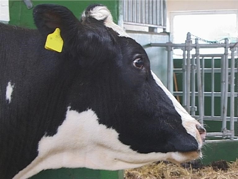
While staring at visitors or attendants, BSE cases typically appear disturbed and anxious and may raise or lower their heads alternately. This may be associated with threatening movements such as head butting, charging, pawing or stamping at the ground and/ or snorting. These behaviours are often referred to as ‘defensive aggression’ because they usually only occur upon approach and when animals are confined and cannot flee away from visitors. Less frequently but very dangerously, BSE cases may show unprovoked aggression towards people.
Video 2: Cow shows aggressive behaviour
This 9-year-old Holstein-Friesian cow responds to a passive observer with pawing of the ground, head tossing and snorting. This aggressive behaviour is elicited by merely standing at the gate without further interference.
BSE cases may keep away from their herd-mates and come last to feed or to the milking parlour, possibly because they have become fearful of their herd-mates or environment. Less frequently, BSE cases may also display unprovoked aggression towards other cattle.
Dullness may be observed when animals are monitored undisturbed over prolonged periods. After some time, BSE cases may settle down and appear dull and inactive in comparison with herd-mates, they may also spend time facing walls.
Restraint difficulties and panic reactions
The unpredictable kicking and other exaggerated reactions of BSE cases make them difficult and often dangerous to handle. Great caution must be taken to approach BSE cases, and their restraint should not be attempted outside sturdy and safe restraining devices. Unfamiliar surroundings or handling, in particular when accompanied by sudden movements, may provoke ‘panic’ reactions. Animals may also stop, startle, tremble or become nervous when coming to cross a previously familiar line on the ground (doorway, pen threshold, bright light reflection on the ground, drain cover or gutter). They may refuse to proceed over these lines as if there was a major physical obstruction. If they can be forced forward, they may take a giant leap over the perceived obstacle. This problem can usually be overcome by spreading bedding over the offending object to mask it.
Video 3: Cow shows hesitancy
This 7-year-old Holstein-Friesian cow is apprehensive of the drain in front of the pen exit. Although the drain is covered by bedding, the cow is very hesitant to cross the drain, sniffs it anxiously and shivers briefly but eventually leaps over.
BSE cases usually try to evade any approach or physical restraint. When restrained, they may vigorously ‘head butt’, especially when approached or touched on the head and neck.
Hyperaesthesia, ‘BSE series’ and ballistic kicking
The hyper-reactivity of BSE cases to certain environmental clues or external stimuli is often described under the term ‘hyperaesthesia’. This may be due to one or more nervous system lesions resulting in hyperaesthesia, hyperreflexia or a disinhibition of lower motor systems. Suspect cases may suddenly back away from any gentle hand approach. The exaggerated events often seen in BSE cases are listed in Table 1. These prolonged and magnified responses may be quantified during routine testing of the function of the cranial nerves.
‘BSE events’ or ‘BSE series of events’
BSE cases may show an increased frequency of one or more of the events listed in this table, either spontaneously or triggered by stress or external stimuli.
‘BSE events’ (may occur spontaneously or be provoked by an stimulus that was previously insignificant to the animal)
excessive and asymmetrical ear movements
head and neck shaking, head tossing, head bobbing
sneezing, snorting
excessive nose licking, nose wrinkling, yawning or flehmen
tooth grinding (bruxism)
tremors
agitated behaviour
excessive vocalisation
unusual hiding, going away in familiar circumstances, including going to face a wall or a corner
over-reaction to visual or auditory stimuli that are part of the normal routine and insignificant to other cattle
sudden jerking movements of limbs, body parts or whole body (startle)
Events mostly seen when external stimulus is applied
startle (sudden jerk of the head, forequarters or whole body)
‘pawing at the ground’ and blowing air forcefully while facing an observer
violent kicking
aberrant startle (drop attacks?): advanced BSE cases may fall over as they startle
‘BSE series of events'
Any series of the events occurring in a close temporal combination is most suggestive of BSE, especially if it can be induced in a repeatable fashion by an external stimulus.
Tests of hyper-reactivity
Tactile stimuli and stick test
Exaggerated responses to tactile stimuli (e.g. head tossing) are usually severe when the head or neck is touched and decrease caudally (see clip below). These responses are often elicited by purely standing in front of an animal that is restrained in a crush. Stroking of the tail head may have a calming effect4
Video 4: Cow shows over-reaction
This 8-year-old Holstein-Friesian cow over-reacts to tactile stimuli when either touched on the head or lightly pricked on the neck with artery forceps, which is characterised by head tossing or head bobbing. She kicks out when pressure is applied on the thoracic spinal column but stays calm when the examiner stands behind her to measure the rectal temperature. Touching the distal part of the hind limbs with a flexible stick results occasionally in quick, forceful kicks.
Some BSE cases show quick and forceful ‘ballistic’ kicking of the hind limbs, which is sometimes repetitive and often leads to early drying off. Touching the hind limbs of BSE suspects should be avoided, unless using a long & flexible stick. This ballistic kicking may occur spontaneously, especially when cattle are being approached from the side or from behind. This reaction may be tested with a flexible plastic (‘PVC’) stick to apply a light touch on the lower hind limbs (‘stick test’). In BSE, this may induce ‘ballistic’ kicking of the stimulated limb.
Video 5: Cow's response to stick test
Repeated touching of the distal parts of the hind limbs with a flexible stick elicits forceful kicks in this 7-year-old Holstein-Friesian cow. It is important to repeat the stick test several times to assess if the animal habituates to the stimulus
By contrast, normal cattle may show no reaction to this test, or they may kick on the first stimulus (usually not forcefully) and stop kicking after the second or third stimulus. Given the potential for serious backlash injuries to the animal or handlers during kicking, we strongly advise against using an inflexible hard stick device (broomstick or other) for this test.
Flash test, bang test or hand clap and clipboard test
Startle reactions are common in BSE cases and may be defined as an exaggerated motor response to unexpected visual, tactile or auditory stimuli. This may take the form of a flinch or jerk of the head, forequarters or of the whole body. Episodically, BSE cases may show sudden jerking of a limbs, head or whole-body startle in the absence of any apparent stimulus (unprovoked startle response).
Video 6: Cow shows spontaneous startle responses
When released into the yard this 8-year-old Holstein-Friesian cow startles several times without obvious stimulus (spontaneous startle responses).
Most typically, many BSE cases will repeatedly startle in responses to visual, tactile or auditory stimuli 13;43. Startle responses may be evoked by sudden loud noises (‘hand clap’ or ‘bang test’), sudden lighting changes (‘flash test’), tactile stimuli or abrupt movements such as a brisk hand approach towards the face or a waving of a clipboard 43. They typically show no habituation to a repeated stimulus. This is in contrast with most normal cattle, which usually will quickly become accustomed to most stimuli after an initial mild flinching or startle response (surprise response).
Video 7: Cow's response to bang test
This 7-year-old Holstein-Friesian cow startles and struggles repeatedly to a sudden metallic sound created by hitting a metal lid with a hammer ('bang'). This test is repeated at least twice since BSE cases usually do not habituate to the stimulus.
Video 8: Cow's response to flash test
A camera flash is held in front of this recumbent 9-year-old Holstein-Friesian cow ('flash test') to test the response to visual stimuli. The cow flinches at every flash, i.e. she does not habituate to the stimulus. Please also note the frequent ear movements after the second flash.
Video 9: Cow shows over-reaction to visual stimuli
BSE cases often over-react to visual stimuli, such as waving a hand towards the animal's face ('approach by hand'). This 7-year-old Holstein-Friesian cow startles initially then tosses her head and snorts towards the moving or later stationary hand.
Video 10: Cow shows startle response
A clipboard with a white sheet of paper attached to it is waved towards two Holstein-Friesian cattle, an 8-year-old cow with BSE and a 7-year-old healthy steer. The cow startles repeatedly whilst the steer stays calm. The test should be repeated to assess if habituation occurs.
Adventitious movements (muscle fasciculations, tremors or myoclonus) or head bobbing and tossing may follow startling events (see ‘BSE series’, Table 1). In extreme cases, startle reactions may culminate in falling, collapsing or seizure episodes.
Other adventitious movements (muscle fasciculations, head bobbing, tremors, myoclonus) and seizures
There may be an increased frequency or excessive duration of superficial muscle fasciculations (of the shoulders, flanks and limbs), tail movements or head and neck shaking or tossing towards the flank. These events may occur in the absence of any obvious exogenous stimuli or be evoked by stress, movements or by insignificant stimuli such as flies, falling feed debris or nearby movement of a person or herd-mate (see ‘BSE series’).
As the disease progress, there may be tremors involving the head and neck, the whole body and/ or the limbs.
Video 11: Cow shows fine head tremor
This 6-year-old Holstein-Friesian cow was filmed shortly after transport. She displays a fine head tremor, which later disappeared when the cow got accustomed to the new environment. Please also note the brief head toss with snort.
Excessive head shaking may take a pathological and stereotypical form resembling the head bobbing seen in some cerebellar lesions. As for muscle fasciculations, tremors and head bobbing may occur spontaneously or they may be triggered or worsened by stress (see ‘BSE series’, Table 1) and by locomotion. Myoclonus and seizures have also been reported, but apparently only in very advanced BSE cases.
Miscellaneous head signs
The following signs have often been attributed to unspecified disturbances of sensory or autonomic cranial pathways.
Excessive nose licking, nose wrinkling, snorting, yawning
BSE cases often display excessive or prolonged episodes of nose licking, nose wrinkling, sneezing, snorting, coughing or yawning, which may occur in single episodes or in repetitive combinations and sometimes together with the other events listed in table 1. Flehmen may also follow such episodes.
Video 12: Cow shows 'BSE series of events'
This 4-year-old Friesian cow displays BSE series of events. Note the nose licking, flehmen and head tossing sideways, once with touching of the flank ('flank licking'). Please also note the flapping of the ears when the cow tosses her head sideways.
Video 13: Cow shows 'BSE series of events' to passive observer
This 4-year-old Friesian cow displays BSE series of events when faced by a passive observer. She tosses her head and licks her nose, which is accompanied by teeth grinding.
Signs that suggest pruritus: head rubbing
Pruritus, alopecia and self-inflicted skin lesions are not as frequent as in scrapie.
Video 14: Cow rubs and nose licks excessively
This 9-year-old Limousin cow rubs her head and neck excessively on the door frame, followed by nose licking and snorting.
Head rubbing may be very vigorous and may cause alopecia or benign skin lesions on the poll region of the head. A positive scratch response is displayed rarely 80.
Teeth grinding (bruxism)
Teeth grinding has been frequently associated with BSE (reported in approximately 40% of cases 19;78). It may occur during BSE series or independently of any triggering stimulus.
Ptyalism
In our experience, spontaneous ptyalism is infrequent in BSE. However, stressed and aggressive BSE cases will very frequently display severe ptyalism upon approach, handling or stimulation.
Posture and locomotion changes
Postural and locomotion disturbances are often mild or absent in early BSE cases. At first, affected cattle may appear ‘stiff’ and refuse to walk or run. Subtle gait deficits may be best evidenced when the animal is made to walk, turn, go over steps or slopes and then run at slow and faster paces.
The more advanced BSE cases may show a wide based stance and a low head carriage.
Video 15: Cow with wide-based stance
This 6-year-old Hereford crossbred suckler cow was ataxic on the hind limbs and displays here a wide-based stance with lowered head.
They may have difficulty getting up, at times showing a period of ‘dog sitting’ in the process 22 (see Fig. 2). In some cases, there is a lumbar kyphosis with dropping of the hindquarters and the hind limbs are placed forward, underneath the abdomen.
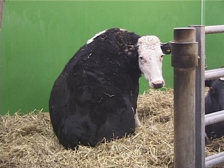
Weakness, paresis and incoordination of the gait (ataxia) are usually first noticed in the hindquarters. There may be lateral swaying and (or) dropping down of the hindquarters and an increased frequency of stumbling, slipping or falling. Pacing has also been observed 11. Knuckling of the fetlocks is an infrequent feature of BSE, which was reported in less than 10% of cases early in the epidemic 78;80. Spasticity, postural tremors and ataxia become more severe as the disease progresses. A mild degree of hypermetria may be present in addition to spasticity.
Video 16: Cow with hind limb ataxia
This 4-year-old Holstein-Friesian cow displays hind limb ataxia (note the swaying of the hip) with mild hypermetria (note the excessive lifting of the hind feet with each step).
Progressive neurogenic gait deficits may eventually lead to recumbency. One or both hind limbs may be stretched out behind in recumbent cattle 54 (Fig. 3).
Recumbent cattle should be considered as BSE suspects unless an alternate cause of recumbency can be clearly established.
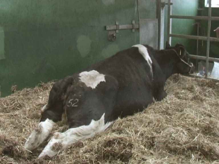
Other historical and physical examination findings
BSE cases typically have normal temperature and mucous membranes. Their respiration rate may be increased when examined in an agitated state. One or more of the following signs may also be noted.
Weight loss and loss of condition
Loss of weight and condition (see Fig. 4) is a frequent feature of BSE and may be its very first sign.
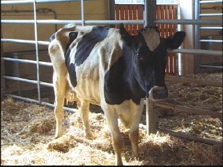
Note the prominent hip bone and the clearly visible rib cage.
Traumatic lesions and / or recumbency
Violent kicking may also result in lesions on the hind limbs.
BSE cases also frequently show traumatic lesions of the skin and deeper tissues, such as lacerations, contusions, haematomata, seroma or other swellings. This may be caused by accidents (slipping, falling or bumping into race sides and fences), which may or may not be witnessed and may result from subtle and otherwise undetected weakness or neurological deficits. Such accidents may also lead to recumbency and inability to stand, often described under the umbrella of ‘downer cow’ syndrome.
BSE should always be considered in the differential diagnosis of unexplained traumatic lesions and recumbency in cattle. This is particularly applicable to abattoirs, where the history of such lesions is not available. With the onset of active surveillance for BSE, it has been shown retrospectively that many of the cases diagnosed in abattoir surveys had displayed signs of BSE that had been overlooked, with presence of swollen joints, falling or recumbency in about 50% of cases 76.
Heart rate and blood pressure changes
Bradycardia and disturbances of the heart rhythm are frequently associated with clinical signs of BSE 3 (see Fig. 5). The most typical finding is that of a low heart rate (60-80 beats per minute) whilst tachycardia would be expected as a result of the animal’s obvious state of stress, fear or excitation 45.
BSE cases may also have an elevated blood pressure 10.
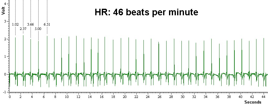
Rumination changes
BSE cases appear to spend as much time eating as normal animals but they appear to spend less time ruminating 2;4. Eructation, swallowing and rumen contractions appear intact in most cases. There have been anecdotal reports of gastro-intestinal ‘bloat’ in BSE cases, which may reflect the average incidence of bloat in cattle.
Milk production
BSE cases may show a decrease in milk yield.
Decreased sensitivity to xylazine
BSE cases may show no or very little reaction to the administration of xylazine in a dose that would induce marked sedation in normal cattle 10.
Ancillary tests
Blood, urine and cerebrospinal fluid analysis
No change has been identified in BSE cases on routine haematology, biochemistry or cytology of the blood, urine or cerebrospinal fluid (CSF) 15;71;72. Despite attempts to find markers of disease that do not rely on detection of the disease-associated prion protein there is currently no pre-clinical test with acceptable sensitivity and specificity to aid in the diagnosis in the live animal. One study failed to identify disease-specific biochemical changes in the pre-clinical stage although it indicated that the energy metabolism of dairy cows incubating BSE was subtly changed 56. Further studies revealed that blood levels of lactic acid and various amino acids were significantly altered in BSE, which was attributed to the exaggerated stress response observed in BSE cases 55.
Changes in the level of selenium, molybdenum and manganese in blood but not in CSF have also been reported in cattle with BSE 38. There have also been attempts to detect a biomarker in the urine of infected cattle 50. The 14-3-3 protein levels in CSF did not appear to distinguish between healthy cattle and cattle displaying early signs of BSE 64. An increased concentration of S-100 protein was found in the CSF of some BSE cases but not in their serum 29;63. Apolipoprotein E and two unidentified proteins were detected in CSF of BSE cases but were absent in healthy controls 41. Others found circulating nucleic acids in BSE-infected cattle, which may be associated with disease 28.
Protein misfolding cyclic amplification (PMCA), which is able to amplify minute amounts of prions in tissues or fluids, has been applied to body fluid samples of BSE-affected cattle: prions were inconsistently identified in saliva and CSF but not in blood 58. A similarly ultrasensitive prion detection method, real-time quaking-induced conversion assay, was used for CSF from experimentally infected cattle with classical and atypical BSE but the results were not consistently positive 53.
In general, blood tests on some of the above and on other components generally struggled to deliver satisfactorily despite earlier promising results 60. Conservation of samples for blood-based tests may also alter test sensitivity or specificity 25. It is likely that the decline in BSE prevalence internationally will prevent the introduction and final development of such tests, if only because test material for development and evaluation will be in limited supply.
Electrodiagnostics
Electrophysiological tests are time-consuming, require special equipment and can only be used on cooperative animals, which is of limited practical value but may be useful for scientific research. Abnormal electroencephalogram 70 and auditory evoked potential recordings 1 were reported in BSE cases but the number of examined cattle was small and included only few controls. Similarly, abnormal retinograms (prolonged b-wave implicit time) were reported in a few cattle with experimentally produced TSEs 57;74. Fluorescence spectra from retinas differed between clinically affected cattle with BSE and BSE-negative cattle 8 but nothing is known about the specificity of an eye-based diagnostic test.
The measurement of parasympathetic activity from electrocardiograms in cattle produced conflicting results regarding diagnostic use 45;51;61.
Differential diagnosis
In the absence of a diagnostic test to confirm BSE in live animals, BSE suspects must be killed to proceed with post mortem diagnosis. In early BSE cases, clear neurological deficits may be absent. Clinical signs may be common to BSE and many other diseases. Diagnosis and/or treatment of other potential causes of illness may take place. Testing and blanket therapy may be indicated, depending on the severity of signs and prognosis for recovery (cost/benefit analysis). Syndromes that share clinical features with BSE may be of extraneural or neural origin. Extraneural diseases include painful conditions, ectoparasitism, slow wasting diseases, ocular diseases and ovarian cysts, which mainly result in weight loss or behavioural changes. A list of the main neurological diseases that may be confused with BSE is provided in Table 2 (see also 7;66;77). One distinctive feature may be the slow onset and the diffuse, symmetrical nature of the neurological deficits in BSE.
Once a suspicion of BSE has arisen, the BSE suspect must be placed under restriction and monitored for a progression of the signs. Restriction is maintained until the animal either fully recovers (from another disease than BSE) or it is killed and tested for BSE.
Table: Neurological diseases that may be confused with BSE
Disorders in bold fit best with the expected age at presentation and clinical signs of BSE, while those followed by an asterisk* are more likely to be encountered in younger animals.
Aetiological category | Diseases | |
Degenerative, developmental disorders, anomalies* | Storage diseases, abiotrophies, etc. Malformations of the central nervous system (including cerebellar aplasia / hypoplasia seen in BVD/MD) & adjacent bony structures* | |
Metabolic, endocrine disorders | Hypomagnesaemia (grass tetany) Hypocalcaemia Nervous ketosis Hepatic encephalopathy | |
Nutritional | Polioencephalomalacia (=cerebrocortical necrosis, Thiamine/ vitamin B1 deficiency)* Copper deficiency | |
Neoplastic, infectious, inflammatory | Brain or spinal cord tumours Viral: Malignant Catarrhal Fever (MCF), rabies, Aujeszky's disease, Tick encephalitis, Borna disease, etc. Bacterial: epidural abscesses (e.g. salmonella), listeriosis Fungal: aspergillosis, cryptococcosis (rare) Parasitic: cysts or migration (sarcocystis, coenuriasis) Other infectious myeloencephalitides (miscellaneous agents) | |
Idiopathic | Brainstem chromatolysis (with hippocampal sclerosis) Cerebral oedema | |
Toxic syndromes | Tetanus, Botulism Miscellaneous plant toxins, mycotoxicoses e.g. Ryegrass staggers Urea poisoning / Ammonia Toxicosis Organophosphate and other insecticide toxicity Mercury poisoning, lead poisoning Salt poisoning | |
Traumatic injuries | Vertebral fractures |
* Diseases that are more likely to be recognised earlier in life than BSE.
Clinical diagnostic criteria
Estimation of BSE prevalence and assessment of the BSE status in a country is largely influenced by the degree of surveillance for BSE. Cattle displaying neurological diseases or cattle with conditions that require emergency slaughter belong to the highest risk group for BSE, and it is important that these cattle are examined for the presence of signs associated with BSE.
Cases reported as suspects
Various clinical procedures have been proposed to detect BSE in live cattle 13;43;59;67. Whilst it is generally acknowledged that cattle that exhibit signs of behavioural, sensory as well as locomotor abnormalities are most likely to have BSE 14;43, the presence of changes in only one or two categories (e.g. an animal that has become nervous without other signs; an ataxic animal that is easy to handle) makes the clinical diagnosis more difficult. In these cases, testing the response to external stimuli (see tests of hyper-reactivity) may be useful to support the clinical suspicion of BSE: cattle with BSE are more likely to respond abnormally to at least two of the four tests of hyper-reactivity 43.
Recumbent and casualty slaughter cattle
BSE may be more difficult to diagnose in recumbent cattle because the gait cannot be evaluated and over-reactivity to external stimuli may not be evident, in particular in end-stage disease where cattle may be dull or stuporous 12;21. In these cases, the clinical history is extremely important because behavioural or locomotor changes may have existed prior to recumbency.
Casualty slaughter cattle often present with recumbency. Any recumbent animal with abnormal positioning of the limbs (see Fig. 3) should be treated as BSE suspect, in particular if they display over-reactivity to external stimuli 49. Over-reactivity was also an important sign in a model proposed to predict BSE in casualty slaughter cattle 39: cattle presenting with at least four of the seven signs aggressiveness, teeth grinding, staring eyes, reduced rumination, recumbency or difficulty getting up and over-reactivity should be regarded as BSE suspects.
Atypical BSE
Atypical BSE cases have been detected world-wide and include countries with low geographical risk of BSE, such as Sweden 27 and the USA 62. The frequency of H- and L-type BSE cases is estimated to be less than 0.5 per million adult cattle tested and both types may represent sporadic diseases 75. Most cases were identified in fallen stock and none were reported as clinical suspects, which suggested that the clinical presentation is unlike classical BSE. Difficulty rising and recumbency but no nervousness or behavioural abnormalities were reported in one Swedish H-type case, which was in late pregnancy and subject to a clinical examination 27. Transmission studies carried out by inoculating cattle intracerebrally with atypical BSE brain homogenate have provided some information on the clinical presentation, which appears to be similar in both H- and L-type BSE. The presence of over-reactivity to external stimuli, unexpected startle responses and ataxia 6;44 resembles classical BSE (nervous form) whereas dullness combined with a low head carriage 6;26;31;44;48;52 (dull form) and compulsive behaviour (licking, chewing, pacing in circles), reported in two experimentally infected cases in the USA 31;57, are usually not associated with classical BSE. Hind limb muscle atrophy was observed in experimentally infected cattle with BASE (L-type BSE) 52. Reluctance to rise or difficulty rising was frequently seen and in the absence of excessive nervousness or over-reactivity may explain why atypical BSE is often diagnosed in downer cows.
Video 17: Steer with low head carriage
This Aberdeen Angus crossbred steer experimentally inoculated with H-type BSE brain homogenate stands motionless with low head carriage in the pen whilst the other cattle are eating (dull form). He also has difficulty getting up.
Video 18: Heifer shows nervous form
This Aberdeen Angus crossbred heifer was experimentally inoculated with L-type BSE brain homogenate and displays the nervous form. She tosses her head towards the hand of an approaching person and responds to touching of the head or approaching with a hood with open-mouthed roaring. There is also loss of coordination, most notable the high-stepping gait (hind limb hypermetria) on trot and gallop.
Further video clips that show the clinical presentation of atypical BSE in cattle can be found in the journals PLoS Pathogens and BMC Veterinary Research article 1 and article 2.
Other prion diseases in cattle
Transmission studies of various strains in cattle have been conducted to inform on the disease phenotype generated by the infection. Although there is currently no evidence that these diseases occur naturally in cattle, it provides some information about the range of clinical signs that can be observed in prion diseases of cattle.
Chronic wasting disease in cattle
Intracerebral inoculation of brain suspension from mule deer naturally affected by chronic wasting disease (CWD) resulted in a disease with relatively unspecific clinical signs: weight loss, inappetence and altered behaviour (apprehension or lethargy, aimless circling) and later recumbency 35. Experimental second passage using brains from affected cattle from the first experiment resulted in similar clinical signs: inappetence, weight loss, dullness, teeth grinding and circling. Although generally unresponsive, cattle reacted to loud noises with exaggerated responses, which included collapsing. All affected animals later became recumbent 37. Similar signs (weight loss, poor appetite, circling and teeth grinding) were also described in cattle infected with elk-derived CWD 34.
Transmissible mink encephalopathy in cattle
Three different inocula containing the transmissible mink encephalopathy agent (TME) agent (Hayward, Blackfoot, Stetsonville isolates) were used to challenge calves intracerebrally and produced a disease characterised by apprehension, inappetence, and decreased water consumption. The display of excitability, over-reactivity to auditory stimuli, teeth grinding and nystagmus was more variable. Hypermetria and hind limb ataxia developed in later stages, followed by recumbency. Although the course of the disease was similar for all three inocula, the rate of clinical deterioration appeared faster with the Blackfoot isolate 65. Another experimental study conducted in the USA several years later to compare first and second passage of TME in cattle by intracerebral inoculation produced a similar disease: hyperexcitability to unfamiliar objects was displayed to various degrees; sudden falling, low head carriage and aimless circling in the pen were also reported 36.
Classical scrapie in cattle
Cattle parenterally inoculated with the classical scrapie agent from sheep or goats (originating from a flock with endemic scrapie in the USA) developed locomotor abnormalities (stilted, stiff gait of the hind limbs, occasionally stumbling and falling and difficulty rising) and dullness with disorientation. Signs usually associated with BSE, such as apprehension or aggressiveness and exaggerated responses to external stimuli, were absent 20. Similar clinical signs were reported for another study in the USA where cattle were intracerebrally inoculated with a pool of brains from scrapie-affected sheep but most cattle also exhibited inappetence and weight loss 24. The clinical presentation did not change on second passage in cattle 23. Other authors reported signs similar to but more subtle than TME in cattle 65.
In the UK, two groups of 10 cattle were intracerebrally inoculated with two temporally separated sources of sheep classical scrapie, which produced again a disease that was mainly characterised by locomotor abnormalities (ataxia with difficulty rising) and dullness (lowered head, leaning against objects) without over-reactivity to external stimuli. Some cattle were also found lying on the side with their limbs paddling. However, two cattle developed a nervous form of the disease with BSE-like signs, such as nervousness and over-reactivity to external stimuli in addition to ataxia 46;47. Video clips that show both disease presentations are available from an article in BMC Research Notes.
Dullness with resting of the head on objects and ataxia, eventually leading to recumbency, was also observed in a calf intracerebrally inoculated with a Spanish classical scrapie isolate 9.
These studies have shown that cattle can develop prion diseases other than BSE, which can be distinguished from BSE by its clinical, pathological and/or biochemical features.
References
1. Arai S, Matsui Y, Fukuda S, Okada H, Onoe S: Brainstem auditory evoked potentials in experimentally-induced bovine spongiform encephalopathy. Res Vet Sci 2009, 87(1):111-114.
2. Austin AR, Hawkins SAC, Kelay NS, Simmons MM: New observations on the clinical signs of BSE and scrapie. In: Transmissible spongiform encephalopathies Proceedings of a consultation on BSE with the Scientific Veterinary Committee of the Commission of the European Communities: 14-15 September 1993; Brussels. edn. Edited by Bradley R, Marchant B. Brussels: European Commission, Agriculture; 1994: 277-287.
3. Austin AR, Pawson L, Meek S, Webster S: Abnormalities of heart rate and rhythm in bovine spongiform encephalopathy. Vet Rec 1997, 141(14):352-357.
4. Austin AR, Simmons MM: Reduced rumination in bovine spongiform encephalopathy and scrapie. Vet Rec 1993, 132(13):324-325.
5. Austin AR, Simmons MM, Wells GAH: Pathological temperament changes in bovines. Ir Vet J 1997, 50(5):304-309.
6. Balkema-Buschmann A, Ziegler U, McIntyre L, Keller M, Hoffmann C, Rogers R, Hills B, Groschup MH: Experimental challenge of cattle with German atypical bovine spongiform encephalopathy (BSE) isolates. J Toxicol Environ Health A 2011, 74(2-4):103-109.
7. Barlow R: Differential diagnosis of bovine neurological disorders. In Pract 1989, 11(2):64-73.
8. Bhattacharjee U, Graham C, Czub S, Dudas S, Rasmussen MA, Casey TA, Petrich JW: Fluorescence spectroscopy of the retina for the screening of bovine spongiform encephalopathy. J Agric Food Chem 2016, 64(1):320-325.
9. Bolea R, Hedman C, López-Pérez Ó, Marín B, Vidal E, Pumarola M, Corbière F, Romero A, Moreno B, Martín-Burriel I, Andréoletti O, Badiola JJ: Experimental transmission to a calf of an isolate of Spanish classical scrapie. J Gen Virol 2017, 98(10):2628-2634.
10. Braun U, Abgottspon S, Gubler E, Schweizer T: Decreased sedation by xylazine and high blood pressure in cows with BSE. Vet Rec 1999, 144(26):715-717.
11. Braun U, Gerspach C, Ryhner T, Hauri S: Pacing as a clinical sign in cattle with bovine spongiform encephalopathy. Vet Rec 2004, 155(14):420-422.
12. Braun U, Hauri S, Kihm U, Ehrensperger F: Evaluation des BSE-Checks bei weiteren 53 Kühen mit BSE [Evaluation of a clinical examination specifically designed to detect bovine spongiform encephalopathy]. Tierärztl Prax Ausg Gros 2007, 35:111-116.
13. Braun U, Pusterla N, Schicker E: Bovine spongiform encephalopathy: Diagnostic approach and clinical findings. Comp Cont Educ Pract 1998, 20(10):S270-S278.
14. Braun U, Schicker E, Hörnlimann B: Diagnostic reliability of clinical signs in cows with suspected bovine spongiform encephalopathy. Vet Rec 1998, 143(4):101-105.
15. Braun U, Schicker E, Pusterla N, Schönmann M: Klinische Befunde bei 50 Kühen mit boviner spongiformer Enzephalopathie (BSE) [Clinical findings in 50 cows with bovine spongiform encephalopathy (BSE)]. Berl Münch Tierärztl Wochenschr 1998, 111(1):27-32.
16. Breslin P, McElroy M, Bassett H, Markey B: Vacuolar lesion profile of BSE in the Republic of Ireland. Vet Rec 2006, 159(26):889-890.
17. Bruce ME, Boyle A, Cousens S, McConnell I, Foster J, Goldmann W, Fraser H: Strain characterization of natural sheep scrapie and comparison with BSE. J Gen Virol 2002, 83(Pt 3):695-704.
18. Casalone C, Zanusso G, Acutis P, Ferrari S, Capucci L, Tagliavini F, Monaco S, Caramelli M: Identification of a second bovine amyloidotic spongiform encephalopathy: Molecular similarities with sporadic Creutzfeldt-Jakob disease. Proc Natl Acad Sci USA 2004, 101(9):3065-3070.
19. Cazeau G, Ducrot C, Morignat E, Lauvergne N, Philippe S, Cohen C, Calavas D: Surveillance clinique de l'ESB en France - Étude quantitative des caractéristiques des animaux et des signes cliniques observes en cas de suspicion [Clinical surveillance of BSE in France - Quantitative analysis of animal features and clinical signs]. Rev Méd Vét 2002, 153(12):785-794.
20. Clark WW, Hourrigan JL, Hadlow WJ: Encephalopathy in cattle experimentally infected with the scrapie agent. Am J Vet Res 1995, 56(5):606-612.
21. Collin E: Étude épidémiologique et clinique des cas de suspicions d'ESB en Côtes d'Armor. Bull Group Tech Vét 2000, 9:195-200.
22. Collin E: Troubles nerveux chez des bovins et ESB. Point Vét 2002, 33(229):40-41.
23. Cutlip RC, Miller JM, Lehmkuhl HD: Second passage of a US scrapie agent in cattle. J Comp Pathol 1997, 117(3):271-275.
24. Cutlip RC, Miller JM, Race RE, Jenny AL, Katz JB, Lehmkuhl HD, Debey BM, Robinson MM: Intracerebral transmission of scrapie to cattle. J Infect Dis 1994, 169(4):814-820.
25. EFSA: EFSA Scientific Report on the evaluation of rapid ante mortem BSE test. EFSA J 2006, 95:1-14.
26. Fukuda S, Iwamaru Y, Imamura M, Masujin K, Shimizu Y, Matsuura Y, Shu Y, Kurachi M, Kasai K, Murayama Y, Onoe S, Hagiwara K, Sata T, Mohri S, Yokoyama T, Okada H: Intraspecies transmission of L-type-like bovine spongiform encephalopathy detected in Japan. Microbiol Immunol 2009, 53(12):704-707.
27. Gavier-Widen D, Noremark M, Langeveld JPM, Stack M, Biacabe AG, Vulin J, Chaplin M, Richt JA, Jacobs J, Acin C, Monleon E, Renstrom L, Klingeborn B, Baron TGM: Bovine spongiform encephalopathy in Sweden: an H-type variant. J Vet Diagn Invest 2008, 20(1):2-10.
28. Gordon PM, Schütz E, Beck J, Urnovitz HB, Graham C, Clark R, Dudas S, Czub S, Sensen M, Brenig B, Groschup MH, Church RB, Sensen CW: Disease-specific motifs can be identified in circulating nucleic acids from live elk and cattle infected with transmissible spongiform encephalopathies. Nucleic Acids Res 2009, 37(2):550-556.
29. Green AJE, Jackman R, Marshall TA, Thompson EJ: Increased S-100b in the cerebrospinal fluid of some cattle with bovine spongiform encephalopathy. Vet Rec 1999, 145(4):107-109.
30. Green R, Horrocks C, Wilkinson A, Hawkins SAC, Ryder SJ: Primary isolation of the bovine spongiform encephalopathy agent in mice: agent definition based on a review of 150 transmissions. J Comp Pathol 2005, 132(2-3):117-131.
31. Greenlee JJ, Smith JD, West Greenlee MH, Nicholson EM: Clinical and pathologic features of H-type bovine spongiform encephalopathy associated with E211K prion protein polymorphism. PLoS One 2012, 7(6):e38678.
32. Gubler E, Hilbe M, Ehrensperger F: Lesion profiles and gliosis in the brainstem of 135 Swiss cows with bovine spongiform encephalopathy (BSE). Schweiz Arch Tierheilkd 2007, 149(3):111-122.
33. Haase B, Doherr MG, Seuberlich T, Drögemüller C, Dolf G, Nicken P, Schiebel K, Ziegler U, Groschup MH, Zurbriggen A, Leeb T: PRNP promoter polymorphisms are associated with BSE susceptibility in Swiss and German cattle. BMC Genet 2007, 8:15.
34. Hamir AN, Kehrli ME, Kunkle RA, Greenlee JJ, Nicholson EM, Richt JA, Miller JM, Cutlip RC: Experimental interspecies transmission studies of the transmissible spongiform encephalopathies to cattle. J Vet Diagn Invest 2011, 23(3):407-420.
35. Hamir AN, Kunkle RA, Cutlip RC, Miller JM, O'Rourke KI, Williams ES, Miller MW, Stack MJ, Chaplin MJ, Richt JA: Experimental transmission of chronic wasting disease agent from mule deer to cattle by the intracerebral route. J Vet Diagn Invest 2005, 17(3):276-281.
36. Hamir AN, Kunkle RA, Miller JM, Bartz JC, Richt JA: First and second cattle passage of transmissible mink encephalopathy by intracerebral inoculation. Vet Pathol 2006, 43(2):118-126.
37. Hamir AN, Kunkle RA, Miller JM, Greenlee JJ, Richt JA: Experimental second passage of chronic wasting disease (CWD(mule deer)) agent to cattle. J Comp Pathol 2006, 134(1):63-69.
38. Hesketh S, Sassoon J, Knight R, Hopkins J, Brown DR: Elevated manganese levels in blood and central nervous system occur before onset of clinical signs in scrapie and bovine spongiform encephalopathy. J Anim Sci 2007, 85(6):1596-1609.
39. Hett AR, Rüfenacht J, Perler L, Heim D: Eine Methode zur Erkennnung von BSE-Verdachtsfällen in der Gruppe der krankgeschlachteten Rinder während der ante mortem Untersuchung [A method to identify BSE suspects in emergency slaughtered cattle during ante mortem examination]. Schweiz Arch Tierheilkd 2002, 144(12):654-662.
40. Jacobs JG, Langeveld JPM, Biacabé A-G, Acutis PL, Polak MP, Gavier-Widen D, Buschmann A, Caramelli M, Casalone C, Mazza M, Groschup M, Erkens JHF, Davidse A, van Zijderveld FG, Baron T: Molecular discrimination of atypical bovine spongiform encephalopathy strains from a geographical region spanning a wide area in Europe. J Clin Microbiol 2007, 45(6):1821-1829.
41. Jones V, Martin TC, Keyes P, Dawson M: Protein markers in cerebrospinal fluid from BSE-affected cattle. Vet Rec 1996, 139(15):360-363.
42. Juling K, Schwarzenbacher H, Williams JL, Fries R: A major genetic component of BSE susceptibility. BMC Biol 2006, 4:33.
43. Konold T, Bone G, Ryder S, Hawkins SAC, Courtin F, Berthelin-Baker C: Clinical findings in 78 suspected cases of bovine spongiform encephalopathy in Great Britain. Vet Rec 2004, 155(21):659-666.
44. Konold T, Bone GE, Clifford D, Chaplin MJ, Cawthraw S, Stack MJ, Simmons MM: Experimental H-type and L-type bovine spongiform encephalopathy in cattle: observation of two clinical syndromes and diagnostic challenges. BMC Vet Res 2012, 8:22.
45. Konold T, Bone GE, Simmons MM: Time and frequency domain analysis of heart rate variability in cattle affected by bovine spongiform encephalopathy. BMC Res Notes 2011, 4(1):259.
46. Konold T, Lee YH, Stack MJ, Horrocks C, Green RB, Chaplin M, Simmons MM, Hawkins SA, Lockey R, Spiropoulos J, Wilesmith JW, Wells GA: Different prion disease phenotypes result from inoculation of cattle with two temporally separated sources of sheep scrapie from Great Britain. BMC Vet Res 2006, 2:31.
47. Konold T, Nonno R, Spiropoulos J, Chaplin MJ, Stack MJ, Hawkins SA, Cawthraw S, Wilesmith JW, Wells GA, Agrimi U, Di Bari MA, Andreoletti O, Espinosa JC, Aguilar-Calvo P, Torres JM: Further characterisation of transmissible spongiform encephalopathy phenotypes after inoculation of cattle with two temporally separated sources of sheep scrapie from Great Britain. BMC Res Notes 2015, 8:312.
48. Konold T, Phelan LJ, Clifford D, Chaplin MJ, Cawthraw S, Stack MJ, Simmons MM: The pathological and molecular but not clinical phenotypes are maintained after second passage of experimental atypical bovine spongiform encephalopathy in cattle. BMC Vet Res 2014, 10:243.
49. Konold T, Sivam SK, Ryan J, Gubbins S, Laven R, Howe MJ: Analysis of clinical signs associated with bovine spongiform encephalopathy in casualty slaughter cattle. Vet J 2006, 171(3):438-444.
50. Lamoureux L, Simon SL, Plews M, Stobart M, Groschup M, Czub S, Graham C, Knox JD: Analysis of clusterin glycoforms in the urine of BSE-infected Fleckvieh-Simmental cows. J Toxicol Environ Health A 2011, 74(2-4):138-145.
51. Little CJ, Julu PO, Hansen S, Mellor DJ, Milne MH, Barrett DC: Measurement of cardiac vagal tone in cattle: a possible aid to the diagnosis of BSE. Vet Rec 1996, 139(21):527-528.
52. Lombardi G, Casalone C, D' Angelo A, Gelmetti D, Torcoli G, Barbieri I, Corona C, Fasoli E, Farinazzo A, Fiorini M, Gelati M, Iulini B, Tagliavini F, Ferrari S, Caramelli M, Monaco S, Capucci L, Zanusso G: Intraspecies transmission of BASE induces clinical dullness and amyotrophic changes. PLoS Pathog 2008, 4(5):e1000075.
53. Masujin K, Orru CD, Miyazawa K, Groveman BR, Raymond LD, Hughson AG, Caughey B: Detection of atypical H-type bovine spongiform encephalopathy and discrimination of bovine prion strains by real-time quaking-induced conversion. J Clin Microbiol 2016, 54(3):676-686.
54. McElroy MC, Weavers ED: Clinical presentation of bovine spongiform encephalopathy in the Republic of Ireland. Vet Rec 2001, 149(24):747-748.
55. Moorby JM, Begley P, Nash RJ, Theodorou MK, Austin AR: Plasma metabolites indicate energy metabolism disruption during the preclinical phase of bovine spongiform encephalopathy infection. Res Vet Sci 2002, 73(2):191-193.
56. Moorby JM, Dhanoa MS, Austin AR: Aspects of the metabolism of dairy cows during the incubation of bovine spongiform encephalopathy. Vet Rec 2000, 147(15):409-412.
57. Moore SJ, West Greenlee MH, Smith JD, Vrentas CE, Nicholson EM, Greenlee JJ: A comparison of classical and H-type bovine spongiform encephalopathy associated with E211K prion protein polymorphism in wild-type and EK211 cattle following intracranial inoculation. Front Vet Sci 2016, 3:78.
58. Murayama Y, Yoshioka M, Masujin K, Okada H, Iwamaru Y, Imamura M, Matsuura Y, Fukuda S, Onoe S, Yokoyama T, Mohri S: Sulfated dextrans enhance in vitro amplification of bovine spongiform encephalopathy PrPSc and enable ultrasensitive detection of bovine PrPSc. PLoS One 2010, 5(10):e13152.
59. Nowotni A, Wendel H, Klee W: Klinische Untersuchung von Rindern auf BSE an einem Vieh- und Schlachthof. [Pre-slaughtering screening of cattle for clinical signs of BSE]. Dtsch Tierärztl Wochenschr 2004, 111(1):5-7.
60. Parveen I, Moorby J, Allison G, Jackman R: The use of non-prion biomarkers for the diagnosis of Transmissible Spongiform Encephalopathies in the live animal. Vet Res 2005, 36(5-6):665-683.
61. Pomfrett CJD, Glover DG, Bollen BG, Pollard BJ: Perturbation of heart rate variability in cattle fed BSE-infected material. Vet Rec 2004, 154(22):687-691.
62. Richt JA, Kunkle RA, Alt D, Nicholson EM, Hamir AN, Czub S, Kluge J, Davis AJ, Hall SM: Identification and characterization of two bovine spongiform encephalopathy cases diagnosed in the United States. J Vet Diagn Invest 2007, 19(2):142-154.
63. Riond JL, Hartmann P, Joller-Jemelka HI, Braun U: S-100 and bovine spongiform encephalopathy. Vet Rec 1999, 145(4):114-115.
64. Robey WG, Jackson R, Walters RL, Brackett JM, Harrington CA, Killian WR: Use of cerebrospinal fluid levels of 14-3-3 in predicting neurodegeneration in confirmed BSE symptomatic cattle. Vet Rec 1998, 143(2):50-51.
65. Robinson MM, Hadlow WJ, Knowles DP, Huff TP, Lacy PA, Marsh RF, Gorham JR: Experimental infection of cattle with the agents of transmissible mink encephalopathy and scrapie. J Comp Pathol 1995, 113(3):241-251.
66. Saegerman C, Claes L, Dewaele A, Desmecht D, Rollin F, Hamoir J, Gustin P, Czaplicki G, Bughin J, Wullepit J, Laureyns J, Roels S, Berkvens D, Vanopdenbosch E, Thiry E: Differential diagnosis of neurologically expressed disorders in Western European cattle. Rev Sci Tech 2003, 22(1):83-82.
67. Saegerman C, Speybroeck N, Roels S, Vanopdenbosch E, Thiry E, Berkvens D: Decision support tools for clinical diagnosis of disease in cows with suspected bovine spongiform encephalopathy. J Clin Microbiol 2004, 42(1):172-178.
68. Saunders GC, Griffiths PC, Cawthraw S, Tout AC, Wiener P, Woolliams JA, Williams JL, Windl O: Polymorphisms of the prion protein gene coding region in born-after-the-reinforced-ban (BARB) bovine spongiform encephalopathy cattle in Great Britain. J Gen Virol 2007, 88(4):1374-1378.
69. Sauter-Louis C, Clauss M, Chaher E, Klee W, Wichmann HE, Kienzle E: Breed predisposition for BSE: epidemiological evidence in Bavarian cattle. Schweiz Arch Tierheilkd 2006, 148(5):245-250.
70. Scott PR: Detailed neurological examination of suspect cases of bovine spongiform encephalopathy (BSE). FRCVS thesis. University of Edinburgh; 1990.
71. Scott PR: Indications for lumbosacral cerebrospinal fluid collection in ruminant species in field situations. Agri Pract 1996, 17:30-34.
72. Scott PR, Aldridge BM, Clarke M, Will RG: Cerebrospinal fluid studies in normal cows and cases of bovine spongiform encephalopathy. Br Vet J 1990, 146(1):88-90.
73. Simmons MM, Harris P, Jeffrey M, Meek SC, Blamire IWH, Wells GAH: BSE in Great Britain: Consistency of the neurohistopathological findings in two random annual samples of clinically suspect cases. Vet Rec 1996, 138(8):175-177.
74. Smith JD, Greenlee JJ, Hamir AN, Richt JA, Greenlee MH: Retinal function and morphology are altered in cattle infected with the prion disease transmissible mink encephalopathy. Vet Pathol 2009, 46(5):810-818.
75. Tranulis MA, Benestad SL, Baron T, Kretzschmar H: Atypical prion diseases in humans and animals. Top Curr Chem 2011, 305:23-50.
76. van Wuijckhuise L, Vellema P, Terbijhe RJ: BSE: klinische diagnostiek en veldervaringen [BSE: clinical diagnosis and field experience.]. Tijdschr Diergeneeskd 2001, 126(8):279-281.
77. Wells GA, Sayers AR, Wilesmith JW: Clinical and epidemiological correlates of the neurohistology of cases of histologically unconfirmed, clinically suspect bovine spongiform encephalopathy. Vet Rec 1995, 136(9):211-216.
78. Wilesmith JW, Hoinville LJ, Ryan JB, Sayers AR: Bovine spongiform encephalopathy: aspects of the clinical picture and analyses of possible changes 1986-1990. Vet Rec 1992, 130(10):197-201.
79. Wilesmith JW, Ryan JBM, Arnold ME, Stevenson MA, Burke PJ: Descriptive epidemiological features of cases of bovine spongiform encephalopathy born after July 31, 1996 in Great Britain. Vet Rec 2010, 167(8):279-286.
80. Wilesmith JW, Wells GA, Cranwell MP, Ryan JB: Bovine spongiform encephalopathy: epidemiological studies. Vet Rec 1988, 123(25):638-644.